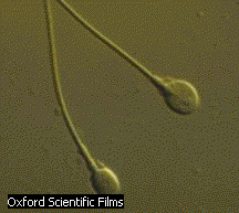
 sperm
sperm
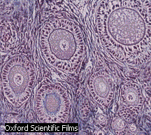 eggs
eggs03/07/2008
Cells
Humans are composed of about 100 trillion cells, with the brain alone having over 100 billion cells - close to the number of stars in the Milky Way galaxy. Just think - all that starts from 1 cell - a fertilized egg. The egg and sperm, also known as germ cells, contain half the number of chromosomes (23) as in a body cell (23x2), also called a somatic cell.
sperm
eggs
To a first approximation, all the somatic cells have the same DNA. Hence you could theoretically clone an individual from a single somatic cell since it contains all 23 pairs of chromosomes- the full genetic blueprint. You have heard about many different kinds of body cells, some of which are shown below.
| cell type | picture |
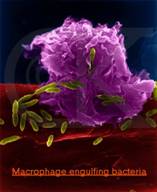
| macrophage
http://www.siue.edu/~cbwilso/MphageEcolli.jpg
|
|
| neuron
http://www2.unil.ch/edab/images/
images_presse/neuron.jpg
|
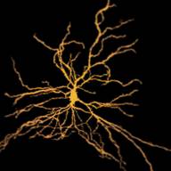 |
| liver (hepatocyte)
http://library.thinkquest.org/
C004535/media/liver_cell.gif
|
 |
| white fat cell
http://www.bioeng.auckland.ac.nz/images/
database/cells/white_adipocyte2.gif
|
 |
| rod
http://www.bioeng.auckland.ac.nz/images/
database/cells/rod_cell2.gif
|
|
| red skeletal
http://www.bioeng.auckland.ac.nz/images/
database/cells/skeletal_muscle_red_fiber.gif
|
 |
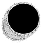
A question should immediately pop to your head. How could so many different kinds of cells be produced from one cell - the fertilized egg, especially since they all have the same DNA. The body cells shown are clearly different from each other and from the starting fertilized egg, shown below with both pronuclei evident.
Somehow, the cells have differentiated from the fertilized egg in the process of development. The fertilized egg is considered to be a totipotent stem cell, since it has the potential to become, through division and differentiation, any kind of cell in the body.
The differentiated cells in the body are different not because their DNA, the carrier of genetic information, is different, but because the DNA blueprint has been read out differently in each cell type. There are about 25-30 thousand different genes in the human genome. In general, a gene is a small part of a chromosome that contains specific information which when decoded produces a specific proteins. Proteins, as we will discuss later, are large organic molecules consisting of 1000's of atoms. Proteins in cells have many functions in immunity, structure, mobility, catalysis, etc. Not all of the genes are read out or expressed (often called turned on). The readout of genes involves the decoding of the information in the DNA sequence into a protein sequence. We will discuss this is more detail later. Muscle cells and neurons differ in that different genes have been turned off and turned on at different times in the development of the cells. Hence the cells, with the same DNA, are composed of different proteins which make the cells look different and have different functions. Proteins are found in all parts of the cell. They are synthesized in the cytoplasm of the cell, but then move to various locations in the cell where they carry out specific functions. Some move to the cell membrane, some to the nucleus, some stay in the cytoplasm, some are secreted from the cell, etc. What controls this process of turning genes on and off?
Genes Expression: Development
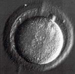
Imagine a fertilized cell. It divides into two daughter cells, which divide into 4 cells, which divide into 8 cells, ..... Are all the resulting cells the same? Have any of the cells become different from the original fertilized egg? (i.e. have any of the cells differentiated?). It turns out that at the 8 cell stage, the developing zygote consists of identical cells. Any one of the cells can be removed and the remaining 7 after insertion into the uterus can lead to the development of a healthy baby. This may seem like a strange experiment, but it has been done for couples that carry a high risk of passing on lethal genetic traits (like Tay-Sachs Disease). The DNA from the 1 removed egg can be analyzed to determine if the cell contains the deleterious gene.
At some point in the process of development of the zygote, the cells do differentiate, and ultimately become liver, skeletal, muscle, neural, blood cells, etc. What happens to cause them to differentiate? At some point, it must come down to the environment of the cells. If you image a large ball of cells, some of them will be on the outside and some on the inside of the ball. They now are clearly in different microenvironments. Somehow the cells sense where they are, and these different environmental signals cause the cells to differentiate. The cells can sense their outer environment, which signals the inside of the cell to turn on or off certain genes, leading cells down the path of differentiation. This process of a cells responding to external environmental cues is call signal transduction.
The DNA of humans and our closest evolutionary relative, the chimpanzee, is 98.7% identical. Most of our genes are identical. How can we be so dissimilar? How does the dissimilarity arise? Paabo et al (Science, 296, pg 340, 2002) compared the genes expressed from 12,000 different genes from different organs (brain, liver, blood) from humans, chimps, macaques, and orangutans. There were little differences in expression of genes in liver and blood. The only big difference was between expression of genes in human brain compared to the rest of the primates.
Gene Expression: Signal Transduction
Cells have to respond to their environment all the time. Immune cells have to respond to the presence of a tumor cell, a bacteria or virus. Retinal cells have to respond to light. Many cells respond to hormones - chemical signals that circulate in the blood. Neurons respond to neurotransmitters - molecules released by adjacent neurons. The responses of the cell can be many: the cell could divide, stop dividing, kill itself, secrete a hormone, take up some nutrients like sugar or vitamins, fire a nerve signal, etc. Problems with the signal transduction can cause big trouble. A cell that should live a normal life span or actually kill itself might live forever and become a tumor cell. A cell that should live might die, which might lead to the manifestations of autoimmune disease (when the body attacks self by mistake) or neurodegenerative disease (like Alzheimer's, Lou Gehrig's Disease, Parkinson's Disease, etc.).
Tumor cells (characterized by unregulated cell growth and cell immortality) are clearly different from normal cells (characterized by regulated growth and a finite cell life). Somehow the normal signaling machinery which puts the brakes on cell growth and division has gone wrong. Normal cells can become tumor cells after enough critical genes have undergone mutations, or in some cases, if the cell is infected with certain viruses that are known to promote cancer. The tumor cell, since it has different properties than the normal cell, must be making different protein than the normal cell (in addition to the proteins that may have been altered through mutation or viral infection).
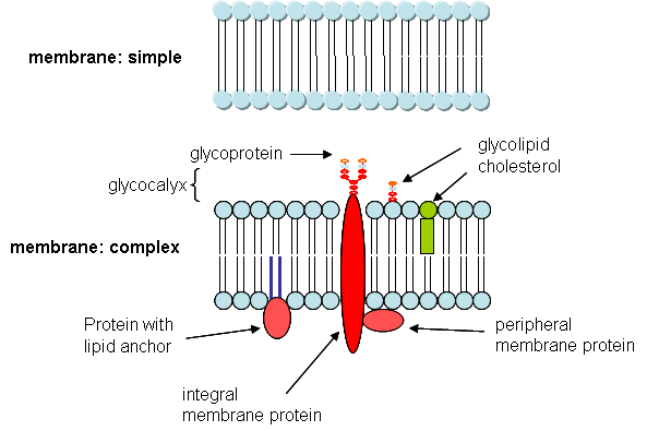
If cells have to respond to their environment, the best place for the cell to place "detectors" to determine what is happening outside of the cell is the cell membrane. These detectors, called receptors, are invariably proteins which span the cell membrane, as shown below.
The outside "signals" are molecules which can bind to the outside or extracellular part of the membrane protein. Signaling starts in several ways:
the protein receptor moves the bound molecule to the inside of the cell. Its presence inside the cell signals the cell to respond. Presumably, the molecule is present in higher concentration outside of the cell than inside or is not found inside the cell at all. The diffusion of molecules from high to low concentration, facilitated by a membrane receptor, is called facilitated diffusion.
the protein receptor changes its shape (conformation) when it binds the outside signal. The shape change also involves the intracellular part of the receptor.
Chemical Modification of Proteins: Kinases and Phosphatases
In most all cases when the outside signaling molecule does not enter the cell, the transmembrane receptor activates protein kinases in the cell. Kinases are a class of enzymes which use a molecule called adenosine triphosphate (ATP) to transfer a phosphate group from ATP to proteins within the cell. molecules. The phosphorylated proteins are either activated or inhibited in the expression of biological activity.
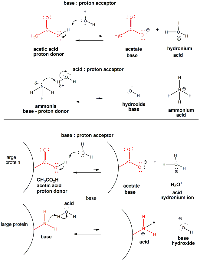
The side chain amino acids of protein are the typical covalent modification of proteins. The simplest of all modifications are those in which amino acids (components of proteins) act as acids or bases to give up or accept protons, in a fashion analogous to the weak acid and base acetic acid and ammonia, as shown below.
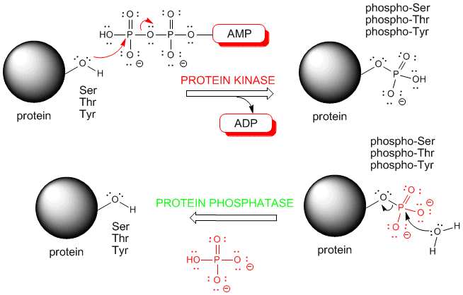
Certain side chains with a ROH group (alcohol) can react covalently with adenosine triphosphate (ATP) in a reaction which transfers a phosphate from ATP to the protein. The enyzme that catalyzes this is a protein kinase.
If a protein is phosphorylated by a kinase, the phosphate group must eventually be removed by another enzyme, a phosphatase. If it wasn't, the phosphorylated protein would be in a constant state of either being activated or inhibited. Kinases and phosphatases regulate all aspects of cellular function. Some people estimate that 1-2% of the entire genome may encode kinases and phosphatases. If so, there might be as many as 800 different kinases and phosphatases. These reactions are shown below.
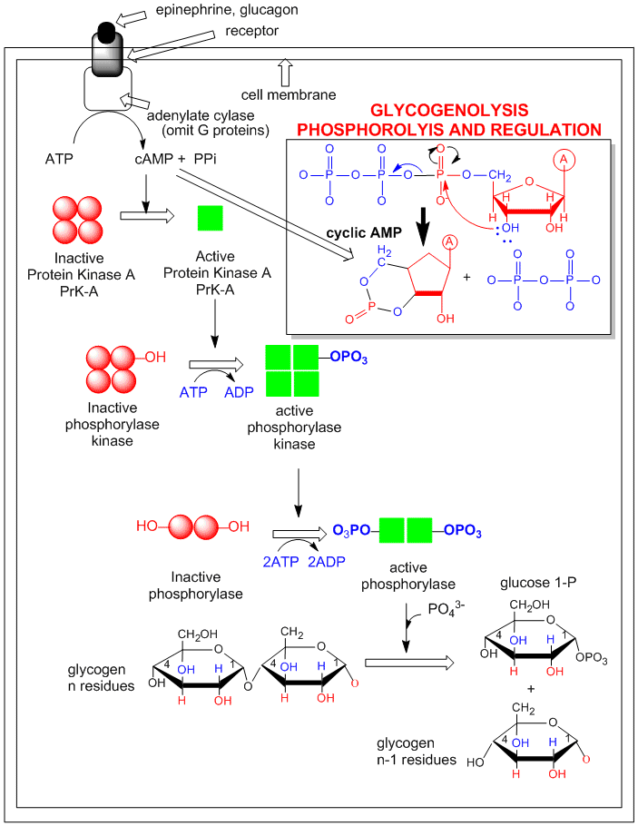
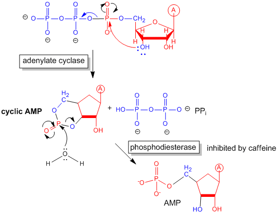
An example of how the hormone epinephrine (a flight/fight hormone) can lead to breakdown of glycogen (your main carbohydrate reserves in muscle and liver) is shown below. The process starts when the hormone epinephrine, released when you sense danger, binds to a receptor in muscle or liver cells. The receptor then changes its shape on the inside of the cell, and bind to and activates another protein enzyme called adenylate cyclase- which chemically converts a small intracellular molecule ATP into an intracellular "second messenger" - cyclic AMP (cAMP). In the cell. cAMP binds to and activates protein kinase A (PKA) (activated by cAMP). which phosphorylates other proteins in the cell. This leads to the activation of the enzyme that breaks down glycogen (glycogen phosphorylase) as shown. The activation mechanism involves a series of kinases, which activate other kinases leading to a cascading amplification of the initial response.
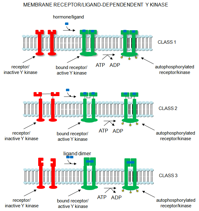
There is a subclass of kinases that have a unique property: when they bind an external signaling molecule on the outside of the cell, the shape change that ensues in their own structure leads them to become active kinases themselves. They don't need a second "messenger" small molecule to be generated in the cell to become active. These receptor kinases often phosphorylate themselves, allowing intracelluar proteins to now interact with them. The target site on the proteins that are phosphoyrlated by these receptor/kinases are tyrosine amino acid present on many target proteins. Many important process like growth and differentiation, and response to signals like insulin are mediated by transmembrane receptor kinase.
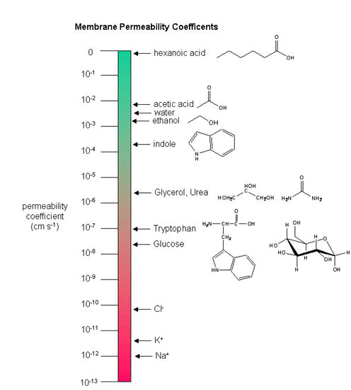
In some cases, the external signal doesn't have to bind to a receptor, it just passes through the membrane of the cell. This occurs in a minor fraction of cases however. Whether a molecule on the outside can pass through the membrane on its own depends on its polarity and size. Think of the barrier a small, charged molecule would face as it tries to cross the nonpolar inner part of the membrane. Remember, ions do not dissolve in nonpolar solvents, as we discovered in lab. In contrast more nonpolar molecules could diffuse through the membrane more readily. Larger molecule have a more difficult time in passing than smaller ones. The figure below shows the permeability coefficient of molecule through a lipid bilayer.
![]()
Quiz: Signal Transduction
