Biochemistry Online: An Approach Based on Chemical Logic

CHAPTER 4 - DNA and the Central Dogma of Biology
A: THE STRUCTURE OF DNA
03/22/16
Note: This chapter on DNA is presented by design short. It is intended to give a cursory overview. More details on nucleic acid structure and its role as the carrier of genetic information can be learned from courses and textbook on Molecular Biology. This guide is intended to give a cursory overview of this complicated field.
A1. The structure of DNA
DNA can exist as single, double-stranded, or mixed forms. It is actually a misnomer to call dsDNA a molecule, since it really consisted of two different, complementary strands held together by IMF's. However, most people talk about a molecule of dsDNA, and so will I. In analogy to protein structure, dsDNA has a linear sequence (primary structure), secondary structure (right handed double helix), and tertiary/quaternary structure (it is folded and packed in the cell).
![]()
![]() Jsmol:
DNA Structure 1
Jsmol:
DNA Structure 1
Alternative DNA structures
The links above show the classic dsDNA helix, in which DNA is in the B form. Other forms of DNA exists, including A-DNA and Z-DNA. Single-stranded sections of DNA can, through intramolecular base-pairing, form stem-loop hairpin structures and quadruplex structures (found at the ends of chromosomes (telomeres).
![]()
![]() Jsmol:
B, A, and Z DNA |
Jsmol:
B, A, and Z DNA |
Structure of a chromosome
Most people have seen pictures of chromosomes viewed through microscopes. Check out this amazing picture of a chromosome taken form Scientific American, September, 1995.
Chromosomes consist of one dsDNA molecule. Each somatic cell of your body has 23 pairs of chromosomes, one member of each pair contributed by your mother and the other by your father. (In germ cells - eggs and sperm - there are 23 individual chromosomes, not chromosome pairs.) One pair are the sex chromosomes, which can come in two forms, X and Y. A pair of X's gives a female, and an XY results in a male.
Figure: Human Chromosomes (with an extra copy of Chromosome 21, which causes Down syndrome)
The human genome has about 3-4 billion base pairs of DNA. (I am uncertain if that estimate is for all 23 pairs, or since each member of a pairs is almost identical, it is an estimate of the DNA in one member each of the just 23 different pairs of chromosomes. I will assume that it is for one member of each pair.) Therefore, on average, each single chromosome of a pair has about 150 million base pairs, which consists of one molecule of DNA and lots of proteins bound to it. dsDNA is a highly charged molecule, and can be view, to a first approximation, as a long polyelectrolyte with a large negative. charge. This very large molecule must somehow be packed into a small nucleus. These packing problem is solved by coiling DNA and packing it with proteins, which usually have a net positive charge. The chromosomes are usually dispersed within the nucleus and are not visible with an ordinary microscope. When the cell is ready to divide, the DNA in the chromosomes replicates, and the chromosomes condense in a fashion that they are not visible using an ordinary microscope. At this point the chromosomes can be stained with a variety of stains (hence the name chromosomes), some of which bind differentially to different chromosomes. The different chromosomes can hence be distinguished by their size, shape, and dye-binding properties, the later called a spectralkaryotype analysis of chromosomes.
The standard picture of a chromosome with which you are familiar, including the one shown above and below, is actually one chromosome of a pair that has just replicated!. One of the chromosomes will stay will the mother cell, and the other will go to the daughter cell. These two chromosomes which are aligned and appear joined at their centers are called sister chromatids. These large DNA/protein complexes must be further packaged in the nucleus, as shown in the "Carl Saganesque" reducing view of the chromosome, a double stranded DNA molecule winds around a core of proteins.
![]() Figure:
Packaging of DNA in the Nucleus
Figure:
Packaging of DNA in the Nucleus
http://www.ncbi.nlm.nih.gov/Class/MLACourse/Original8Hour/Genetics/chromosome.html
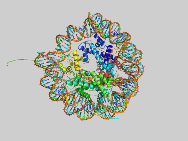
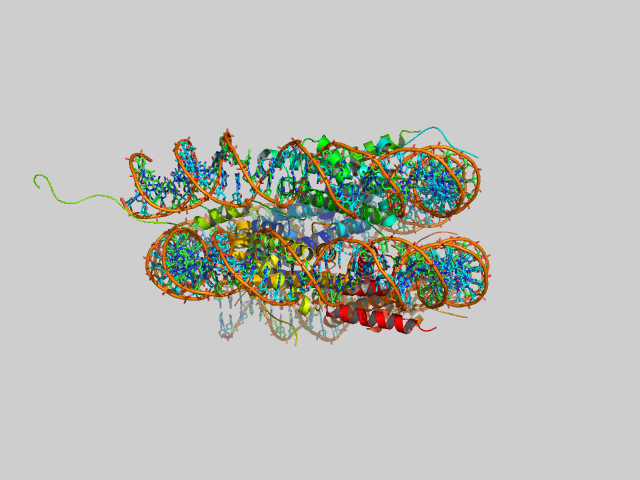
The core is called a nucleosome, and can be viewed under an electron microscope if the chromosome is dispersed. The nucleosome consists of 8 positively charge proteins called histones. In the core are 2 copies each of His 2A, 2B, 3, and 4. The dsDNA winds around the nucleosome core about 2.5 times. The dsDNA then links around other nucleosome in which each nucleosome is connected by a small section of interconnecting spacer DNA, to which is bound another histone, H1. Under an electron microscope, the DNA looks like a bead on a string. The beads are nucleosomes, and the string is the dsDNA.
![]() Figure:
Nucleosome (top and side view)
Figure:
Nucleosome (top and side view)
 Navigation
Return to Biochemistry Online Table of Contents

Biochemistry Online by Henry Jakubowski is licensed under a Creative Commons Attribution-NonCommercial 4.0 International License.