Figure 2from: Baumgart, T., Hess, S., and Webb, W. "Imaging coexisting fluid domains in biomembrane models coupling curvature and tension." Nature. 425, 821-824 (2003)
Reprinted with permission of Nature, and with permission of the authors.
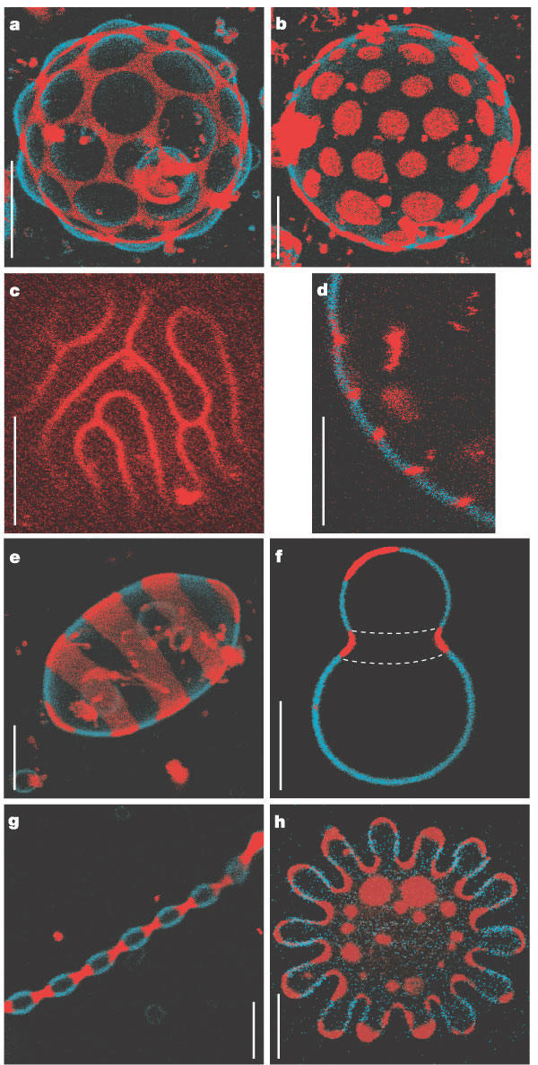
Fluorescence microscopy (two different wavelengths) actual GUVs: Lo phase (blue); Ld phase (red)
2a and 2b: Domains are circular at room temperature and appear to arrange into hexagonal patterns. If the temperature is increased (to point below the two phases mix), the domains undulate laterally. If the temperature is raised further (past the Tm for the phase mixing, the membranes become homogeneous in phase and presumably composition.
c. focus is on top of vesicle, showing only Ld phase
d. equatorial section of vesicle in c
e, f. When vesicles are warmed above the Tm, and then cooled just below the Tm, many small domains develop. The Ld phase is found in the saddle shape regions, and in the tips, both area with high curvature. The Lo phase is found in less curved "tube-like" regions, suggesting its preference for low curvature