Biochemistry Online: An Approach Based on Chemical Logic

 Homework
Problems - Literature Learning Module: Carbohydrates 1
Homework
Problems - Literature Learning Module: Carbohydrates 1
Assessment of Biochemistry/Molecular Biology (BMB) Foundational Concepts
Henry Jakubowski, Ph.D., Professor, Chemistry Department, College of Saint Benedict/Saint John's University
This page contains assessment/exam questions using data, figures, and graphs from research journals such as the Journal of Biological Chemistry which allow their use, or from journals such as from PLOS that are completely open access The papers and topics chosen were selected to assess student understanding of the American Society for Biochemistry and Molecular Biology (ASBMB) foundational concepts and learning objectives as well as MCAT2015 foundational concepts and objectives. These two sets of standards broadly overlap. Both ASBMB and the MCAT2015 strongly emphasis scientific inquiry and reasoning skills, which are perhaps best assessed by open-ended questions derived from the literature in which students must employ higher level Bloom skills of application and analysis.
These questions can also be used by students who seek more opportunities to practice interpreting research literature results. The ability to apply, analyze, and evaluate information and concepts are at the heart of scientific inquiry and reasoning skills which are central to the new ASBMB and MCAT2015 competency standards. The questions in this learning module are designed to assess these competencies using open-ended responses instead of multiple-choice questions. Answers can be found at the link at the bottom of this page.
Research Paper: Trypanosoma brucei Glycoproteins Contain Novel Giant Poly-N-acetyllactosamine Carbohydrate Chains.Abdelmadjid Atrih, Julia M. Richardson, Alan R. Prescott, and Michael A. J. Ferguson. J. Biol. Chem., Vol. 280, Issue 2, 865-871, January 14, 2005
A densely packed layer of a surface glycoprotein covers the surface of the African trypanosome, Trypanosoma brucei. The parasitic protozoa are transmitted by tsetse fly and causes African sleeping sickness in humans and nagana in livestock. In the mammalian host, T. brucei live and divide extracellularly in the blood, lymph, and interstitial fluids. They have 100's of genes for the glycoproteins which produce so many variants that the organism can elude the immune system. The parasite has a deep invagination of the plasma membrane, called the flagellar pocket. Endocytosis and secretion occurs from this site. Little is known about its unique membrane glycoprotein structures and the endosome (vesicles that have undergone endocytosis)/lysosome (degratory vesicles that fuse with lysosomes) system.. Something in the pocket binds the lectin ricin and tomato lectin. Ricin is a heterodimeric protein consisting of a Galβ1-4GlcNAc binding subunit, B chain (32 kDa), and a toxic subunit, A chain (ca. 30 kDa), connected through a disulfide linkage. Tomato lectin is known to bind a linear polymer of N-acetyllactosamine (poly-LacNac), which is comprised of up to 26 Galβ1-4GlcNAc linked principally by 4GlcNAcβ1-3Galβ1- links (which in mammals is found in both the cell and lysosomal membrane glycoproteins). A study was done to exam the flagellar pocket of the bloodstream form of Trypanosoma brucei by studying the ricin-binding glycoprotein.
1. Ricin-binding glycoproteins were purified as follows.
a. Cell were isolated from blood, subjected to hypotonic lysis to release cytosolic components and soluble forms of surface glycoproteins, and cell ghost (cells without cytoplasm and other soluble contents) collected. Briefly explain this procedure and how it works with words and pictures.
b. Cell ghosts were solubilized in 50 mL of 8 M urea, 3% SDS, 50 mM Tris-HCl, pH 6.8. Explain the role of each of the added ingredients. How do they collectively solubilize the cell ghost.
c. The extract was diluted 50 fold 50 mM Tris-HCl, pH 6.8, 0.4 M NaCl, 0.8% Triton X-100, and protease inhibitors. Ricin-coupled agarose chromatography beads were added and gently mixed overnight. The beads were collected by gentle centrifugation and packed into a column. Why was the extract diluted 50 fold before the chromatography beads were added? Why did they switch from SDS to Triton X-100? Draw a diagram showing the ricin beads in the column and any interacting species.
d. Bound proteins were eluted from the column after extensive washing. Without altering pH or ionic strength, how could bounds proteins be eluted? Name the eluting agent.
2. Aliquots of the eluted fractions were separated by PAGE. A membrane was placed on the gel and electrophoresis of the proteins onto the membrane was performed (Western Blot). The results are shown below.
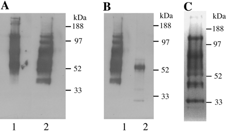
Fig 1. Ricin blot analysis of glycoproteins from bloodstream form T. brucei. Glycoproteins were eluted from a ricin-agarose column. After electrophoresis, glycoproteins were either transferred onto nitrocellulose membrane and probed with horseradish peroxidase-conjugated ricin (A and B) or stained for carbohydrate with periodic acid-Schiff (C). A: lane 1, native extract; lane 2, extract reduced with dithiothreitol (before electrophoresis). B: lane 1, reduced extract; lane 2, reduced extract after digestion with PNGase-F. C: reduced extract. The positions of molecular mass standards are indicated.
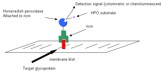
a. Draw a picture showing the electrophoreses protein on the membrane and how it react and is visualized with horseradish peroxidase-conjugated ricin
b. In gel A, explain the chemical effect of dithiothreitol on the proteins which do to alter the migration of the proteins.
b. Why is there only one main band left after treatment with PNGase-F before the electrophoresis. (Hint: Google the enzyme).
![]()
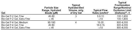
3. Glycans released from the ricin-binding glycoproteins by PNGase-F were separated by Bio-Gel P4 gel filtration chromatography.
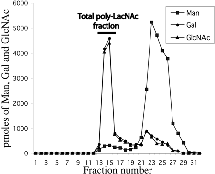
Fig. 2. Bio-Gel P-4 chromatogram of glycans obtained from ricin-binding glycoproteins. Glycans released from ricin-binding glycoproteins by the action of PNGase-F were purified from detergents and salts. The monosaccharide compositions of the fractions were determined by GC-MS. The void volume total poly-LacNAc fraction is indicated with a bar.
Two major fractions were found. Which peak has the highest average molecular weight species? Give an estimate of the molecular weight range of the glycans found in the void volume of the Bio-Gel P-4 column, based on the information from BioRad below.
4. The poly-LacNAc fraction from the Bio-Gel P4 column was chromatographed on a HPLC Dionex CarboPac A-100 high pH anion exchange column. (HPAEC), used for the analysis of weakly ionizable carbohydrates. The beads in this column consist of 7.5-μm diameter vinylbenzyl chloride/divinylbenzene macroporous substrate fully functionalized with an alkyl quaternary ammonium group (15%crosslinked). The column is equilibrated in 95% solution A (100 mM NaOH) and 5% solution B (0.380 M sodium acetate in 0.1 M NaOH) and bound glycans separated using a linear gradient of 5-40% solution B.
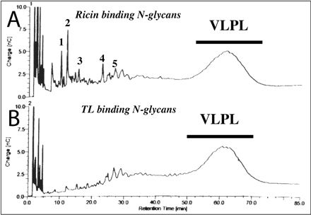
Fig. 3. Dionex HPAEC separation of total poly-LacNAc fractions. A, HPAEC chromatogram of glycans, detected by pulsed-amperiometric detection, of the total poly-LacNAc fraction isolated after Bio-Gel P-4 gel filtration (see Fig. 2). B, HPAEC chromatogram of the glycans of the total poly-LacNAc fraction which bind to tomato lectin-agarose. The VLPL (very large poly LacNac) is indicated with a bar.
What is the pH of the column during equilibration and running? Why was this column run at this pH.
5. The VLPL fraction was subjected to monosaccharide compositional analysis by GC_MS. A 1 ul aliquots that was desalted and dissolved in water was mixed with 1 ul of acetonitrile with 2% formic acid and subjected to GC-MS. .It contained Man, Gal, and GlcNAc in the ration of 3:59:60. Is this consistent with the proposed glycan structure?
6. It is necessary to know the linkage of the monosaccharide in the VLPL. Linkage analysis was done by converting glycans to partially methylated alditol acetates and analyzing them by GC-MS. The general chemistry involved in the formation of methylated alditol acetates used in methylation analysis is shown below. Definitive identification requires, in addition to the analysis of the MS pattern, the comparison of retention times with those of known standards
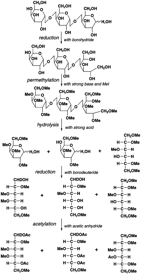
a. Why is methylation of free OH groups by basic CH3I required before hydrolysis of the glycosidic links? What do methyl group denote?
b. Why is the reduction carried out?
c. Why is a final acetylation carried out? What do acetyl groups mark?
"Traditional MS approaches are based on the fragmentation of organic molecules under some kind of impact, followed by the differentiation of the resulting particles according to their mass-to-charge ratio. In methylation analysis, peaks are identified by a combination of their retention time (relative time of elution from the LC column) and their electron impact (EI)-MS fragmentation pattern. The EI fragmentation patterns of similarly substituted monosaccharides of the same class (e.g., aldohexoses) are the same. Thus, definitive identification requires, in addition to the analysis of the MS pattern, the comparison of retention times with those of known standards (i.e., all 2,3,4-tri-O-methyl-hexoses produce the same EI-MS spectrum, but peracetylated 2,3,4-tri-O-methylgalactitol elutes later than peracetylated 2,3,4-tri-O-methylglucitol). This type of analysis indicates which residues are terminal (i.e., they are methylated at every position except OH-1 and OH-5) and how each monosaccharide is substituted; the latter reveals, inter alia, the occurrence of branching points. However, methylation analysis neither indicates which residues are attached to each other (i.e., does not provide sequence information) nor if a particular linkage is of the α or β configuration." NCBI
The methylation analysis in conjunction with NMR analysis showed large amounts of 4-)-substituted-(β)GlcNac and 6-O-substituted-(β)Gal. NMR analysis shows most of the GlcNAc and Gal residues were present as LacNac repeats of approximately 54.
Finally, fluorescein isothiocyanate-labeled ricin was added to fixed tryptosomes and detected by fluorescence microscopy.
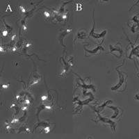
Fig 5. Subcellular localization of ricin-binding glycoproteins in bloodstream form T. brucei by fluorescence microscopy. Fixed trypanosomes were stained with FITC-ricin (A) and FITC-ricin in the presence of the ricin-blocking substances (as a control).
7. What ricin-blocking substances might they have used in panel B?
Answers: Literature Learning Module - Carbohydrates 1
 Navigation
Return to Biochemistry Online Table of Contents

Biochemistry Online by Henry Jakubowski is licensed under a Creative Commons Attribution-NonCommercial 4.0 International License.