Biochemistry Online: An Approach Based on Chemical Logic

Chemistry Online: An Approach Based on Chemical Logic
List of Figures
04/17/16
I have created most of the images found in BC Online, using programs such as Power Point, Paint Shop Pro, and ChemDraw. Some of these figures have been modeled after those found in various biochemistry textbooks. I have also used other figures which are already available on the web. For those, I have referenced those by including the url for the site directly on the figure.
Some of the figures in this online book are used, by permission, from Science, Nature, and the Proceedings of the National Academy of Science. The specific reference for each figure is shown on the figure. For these specific figures:
"Readers may view, browse, and/or download material for temporary copying purposes only, provided these uses are for noncommercial personal purposes. Except as provided by law, this material may not be further reproduced, distributed, transmitted, modified, adapted, performed, displayed, published, or sold in whole or in part, without prior written permission from the publisher."
Introduction
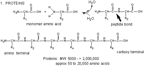
- Amino Acids React to Form Proteins
- Cyclization of simple sugars through hemiacetal links
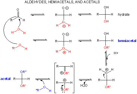
- Hemiacetal/acetal chemistry
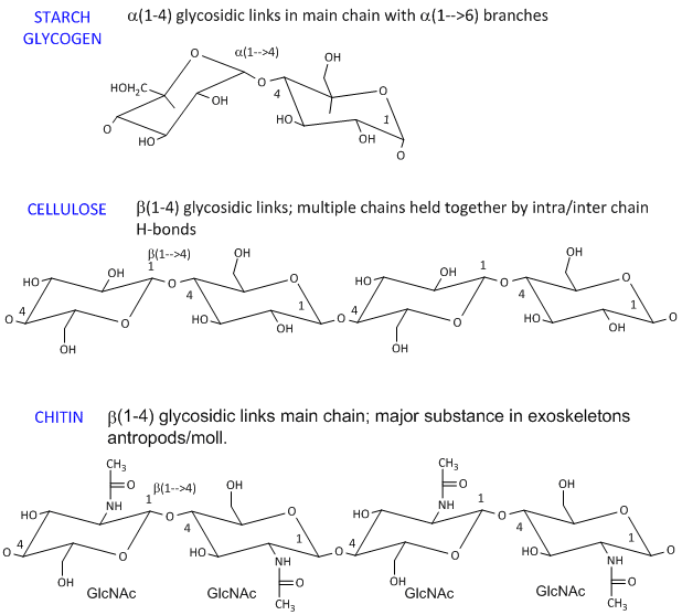
- Examples of complex carbohydrates
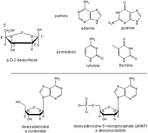
- Nucleic Acid Monomers
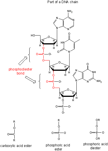
- Nucleic Acid Polymers
- Map and description of course
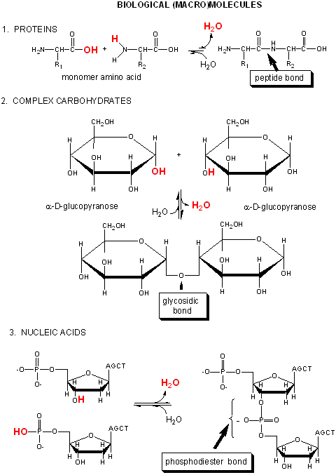
- Comparison of protein, carbohydrate and nucleic acid synthesis
Chapter 1: Lipid Structure
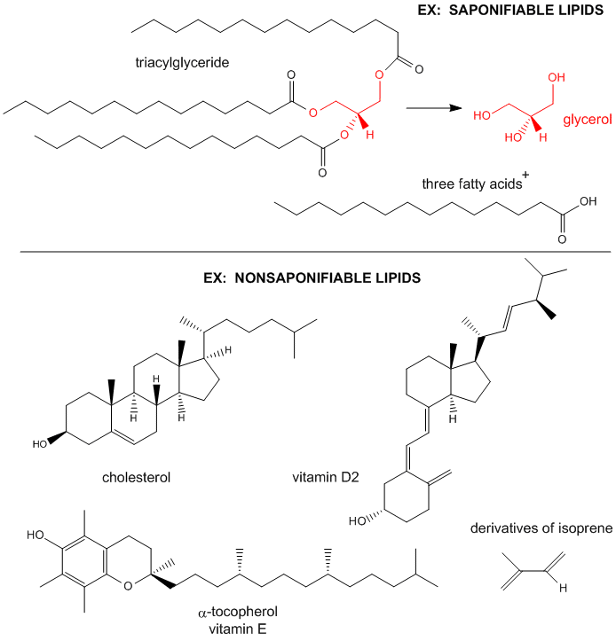
- Examples saponifiable and nonsaponifiable lipids
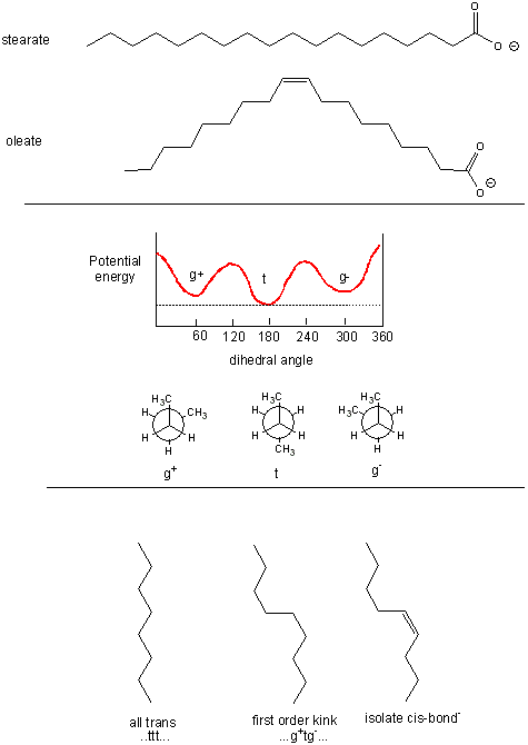
- Conformations of fatty acids
- Classification of common phosopholipids, glycolipids, and triacylglyerides
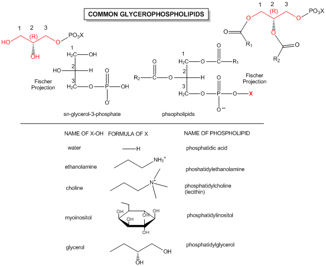
- Structures of common phospholipids
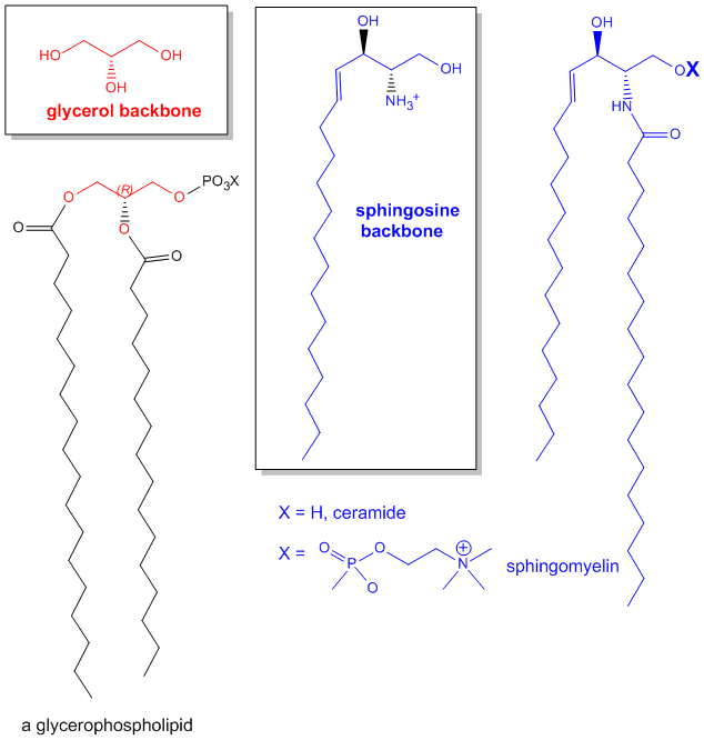
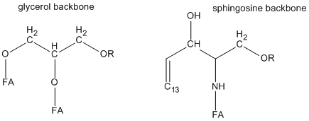
- Comparison of lipids with glycerol and sphingosine as backbones
- Conformations of butane and fatty acids.
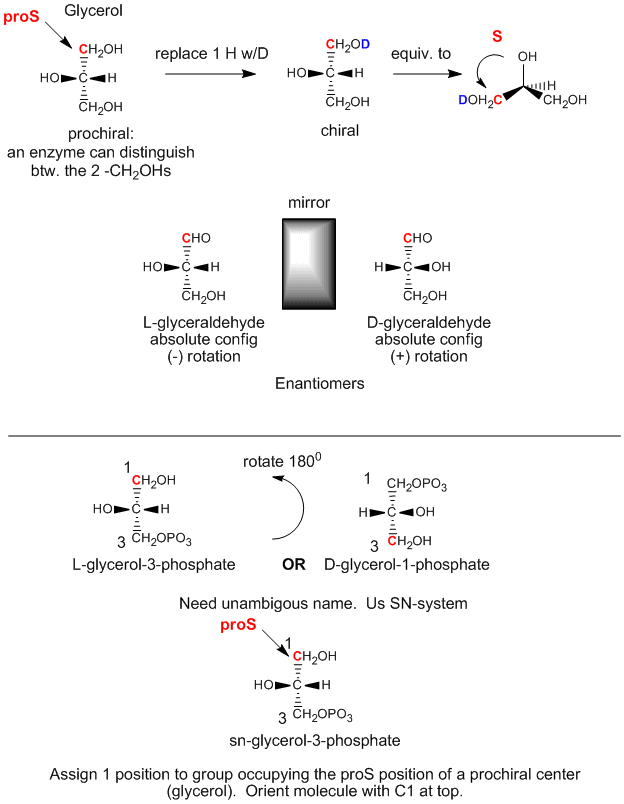
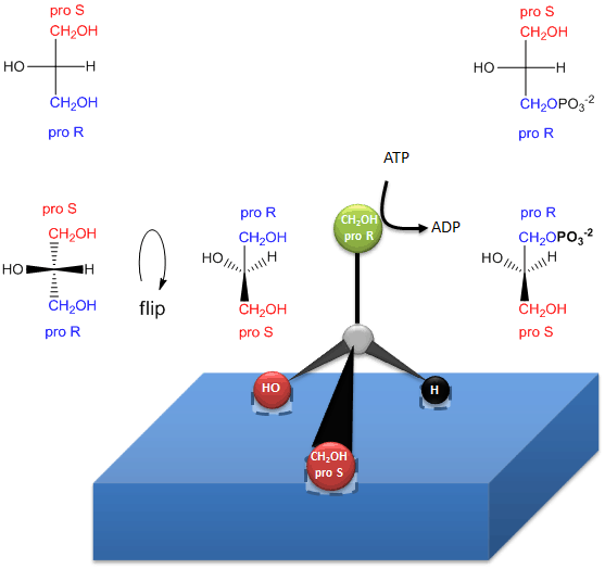
- Glycerol - A prochiral molecule
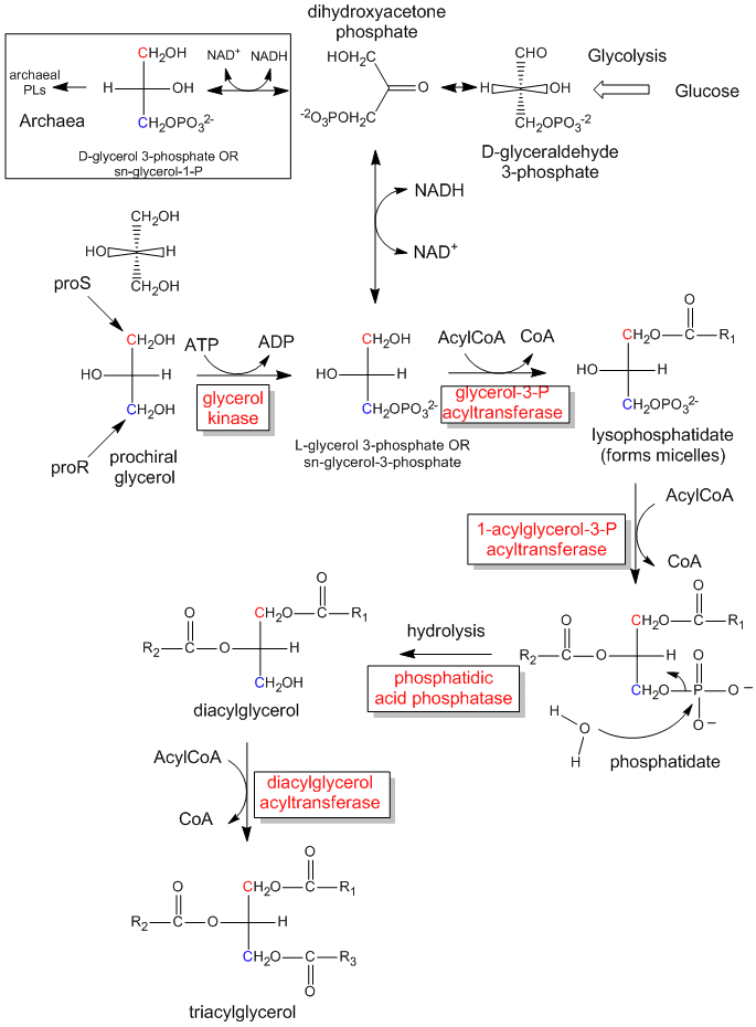
- The biological synthesis of triacylglycerides and phosphatidic acid from prochiral glycerol.
- How an enzyme (glycerol kinase) transfers a PO4 from ATP to only the proR CH2OH of glycerol in formation of chiral triacylglycerols and phosphatidic acid.
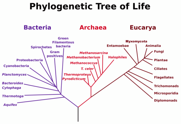
- Phylogenetic Tree of Life
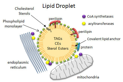
- Lipid Droplets
- A single chain amphiphile jumps into water!
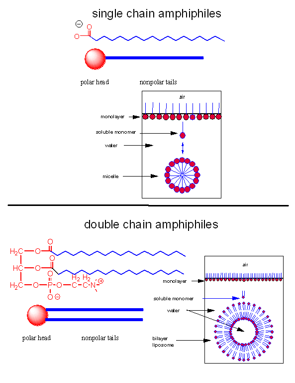
- Structures of single and double chain amphipiles in water - Micelles and Bilayers
- Pentaphenyl fullerene (Ph5C60K) in water.
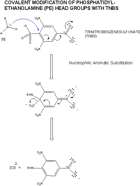
- Nucleophilic aromatic substitution
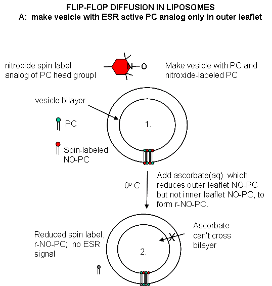
- Flip/Flop diffusion in liposomes A: Making vesicles with ESR active PC analog only in outer leaflet
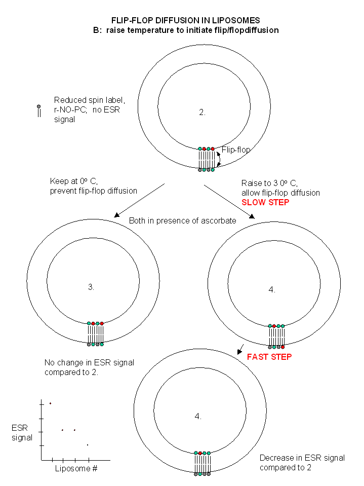
- Flip/Flop diffusion in liposomes B: raising the temperature to initiate flip/flopdiffusion.
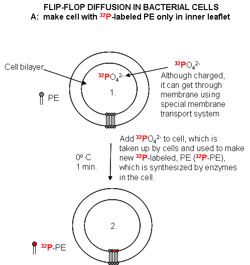
- Flip/Flop diffusion in bacterial cells A: labeling inner leaflet phospholipids with 32 P
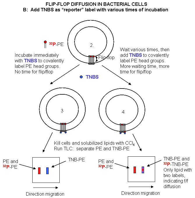
- Flip/Flop diffusion in bacterial cells B: labeling Cells in A with TNBS to detect Flip/Flop
- Comparison of glycerol and sphingosine headgroup structure
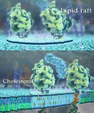
- Lipid Rafts enriched in SM and Cholesterol
- Heat absorption and water: phase transitions
- Phase transition for DPPC (Dipalmitoyl phosphatidylcholine)
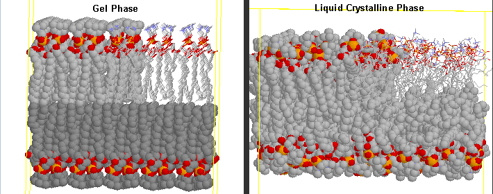
- Differences in Gel and Liquid Crystalline Phases in Phospholipids Bilayers
- TM di16:0 PC and di16:0 PA - Energy Diagram
- Phase Transition Temperatures for glycerophospholipids
- Equatorial cross sections of GUVs with multiple phases
- GUVs structures with multiple phases
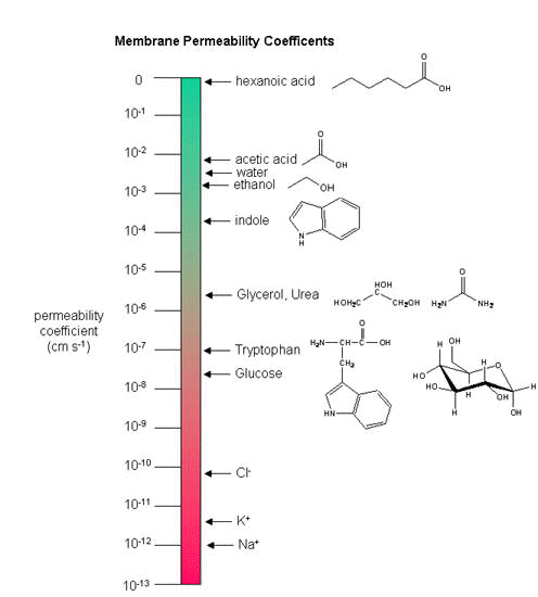
- Permeability of liposome bilayers
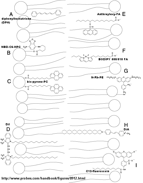
- Fluorescent membrane probes
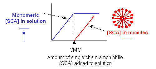
- Graph showings distribution of single chain amphiphiles in bulk aqueous solution
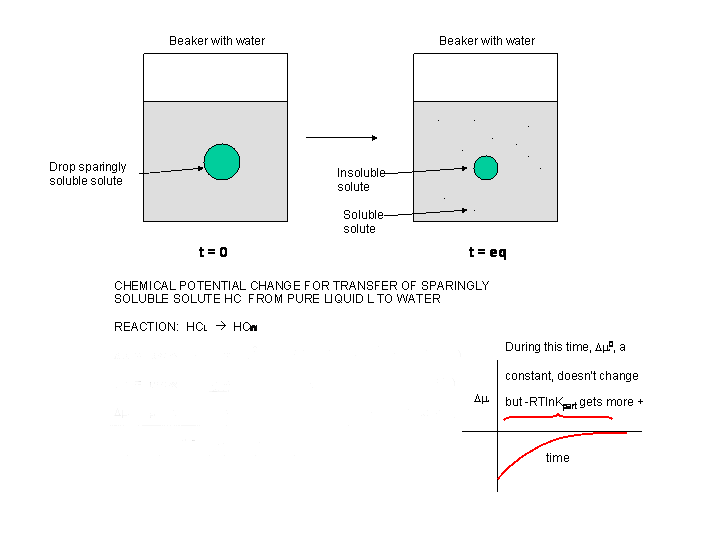
- Δμ for transfer of a sparingly soluble liquid solute into water
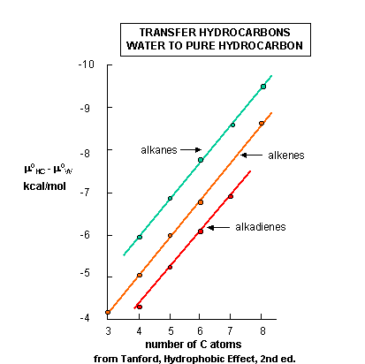
- Standard free energy of transfer of HC from aqueous solution to a pure liquid hydrocarbon.
- Transfer of Aliphatic Alcohols and Fatty Acids from Water to Pure Liquid; Thermodynamic Parameters for Transfer of Aliphatic Alcohols From the Pure Liquid to Water.
- Surface Area per Head Group vs no. of C in Amphiphile - Globular, Cylindrical, Planar Forms
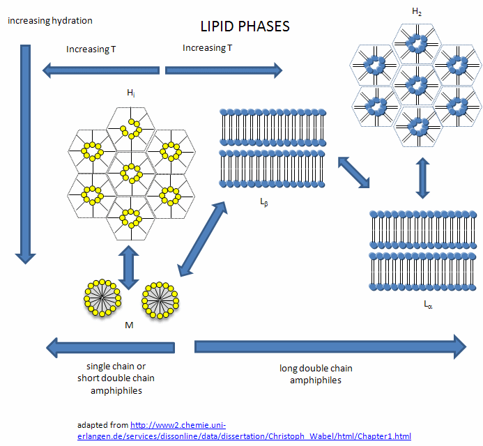
- Lipid Phases
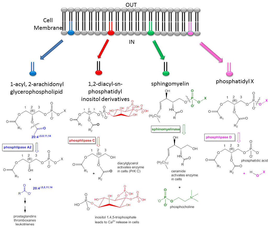
- Lipids - More than Grease - Mediators in Signal Transduction
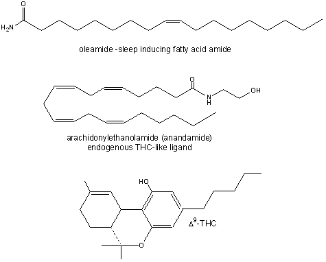
- Fatty Acid Amides: Neurochemical Mediators
Chapter 2: Protein Structure
- Table: Structure and Property of the Naturally-Occurring Amino Acids
- Amino Acids React to Form Proteins
- Review: Comparison of protein, carbohydrate and nucleic acid synthesis
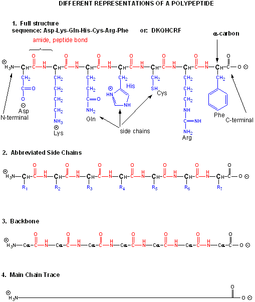
- Different Representations of a Polypeptide (heptapeptide rep)
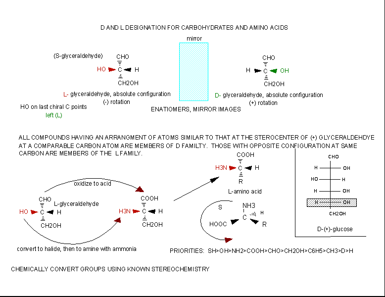
- Stereochemistry of Amino Acids
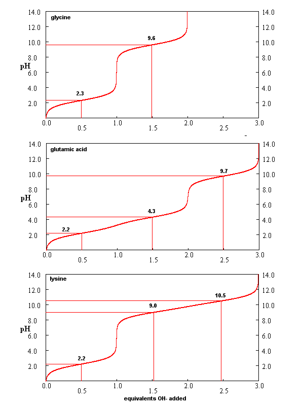
- Titration curves for Gly, Glu, and Lys
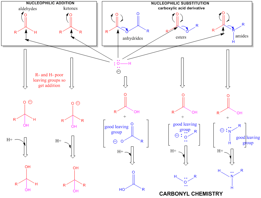
- A review of the chemistry of aldehydes, ketones, and carboxylic acid derivatives
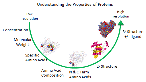
- Understanding the properties of proteins: low to high resolution
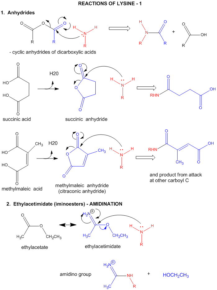
- Lysine Reactions 1
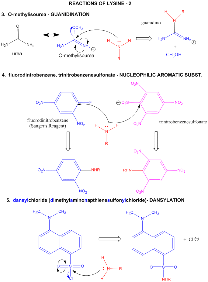
- Lysine Reactions 2
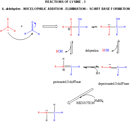
- Lysine Reactions 3
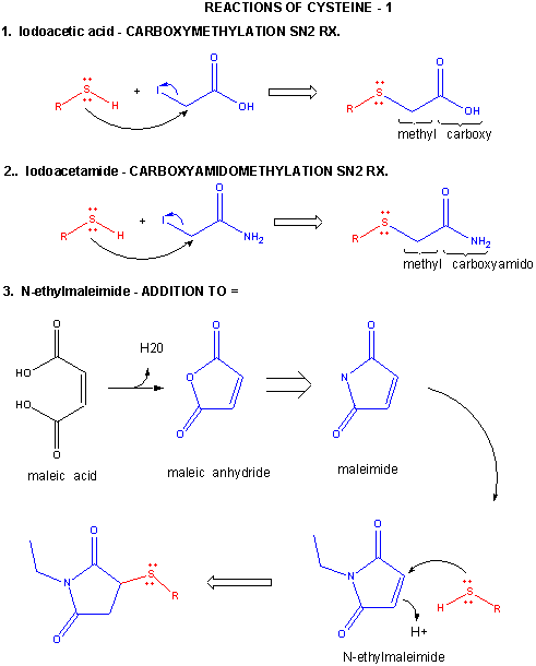
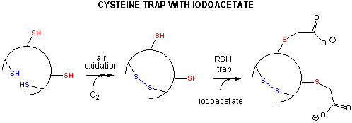
- Cysteine Reacions 1
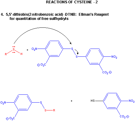
- Cysteine Reactions 2
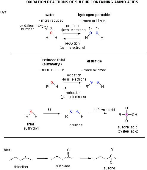
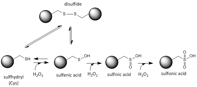
- Sulfur redox chemistry
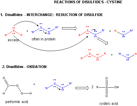
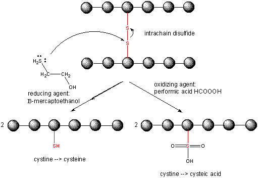
- Disulfide - Cystine - Reactions
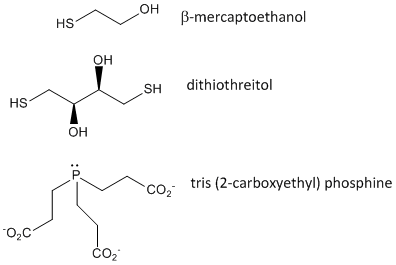
- Disulfide Oxidizing Agents - β-mercaptoethanol, dithiothreitol and phosphines
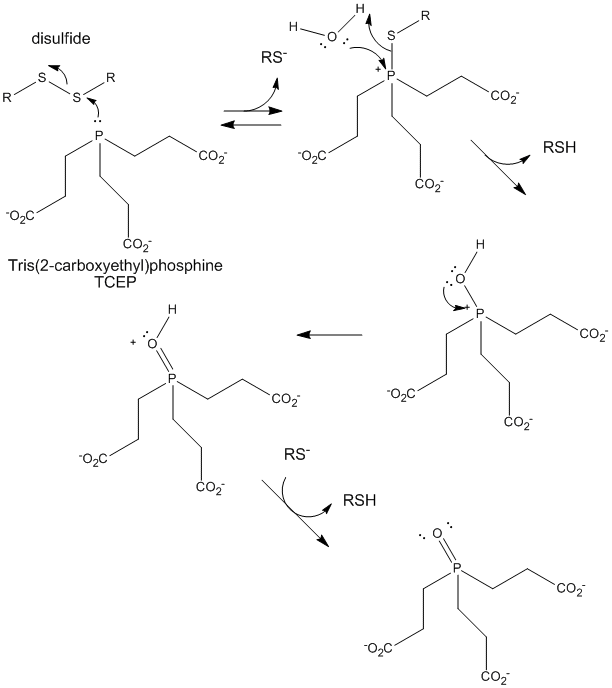
- TCEP reduction of disulfides
- Cleaving Disulfide Bonds in Proteins
- Redox state of Cysteine
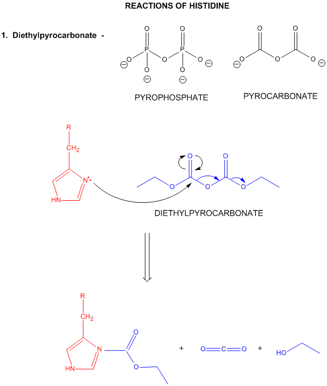
- Reactions of Histidine
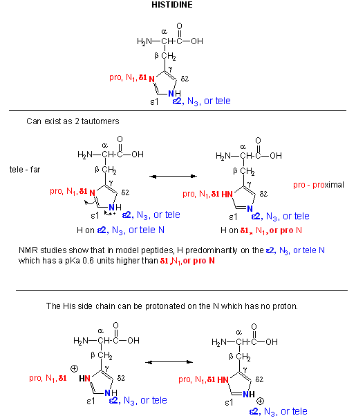
- Where is the H on His? Where is the Charge?
- His reacts with reasonably high selectivity with diethyl pyrocarbonate
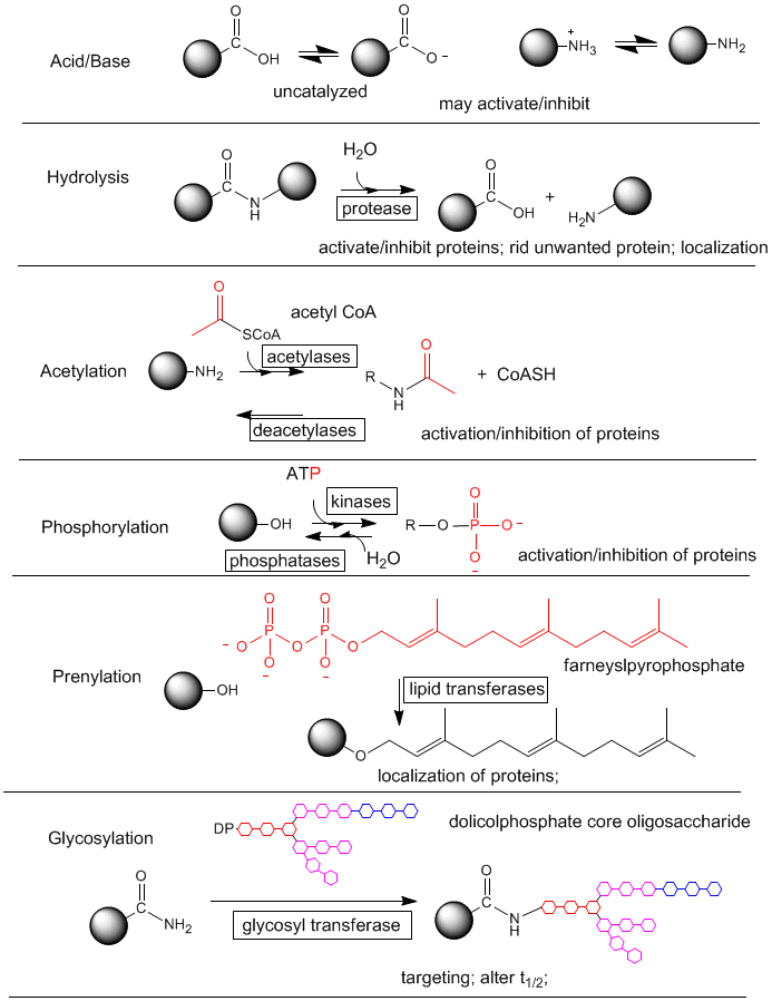
- Post-translational modification of proteins
- Understanding the properties of proteins: low to high resolution
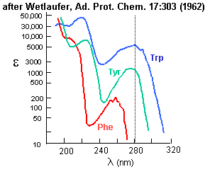
- Amino Acid absorbance profiles.
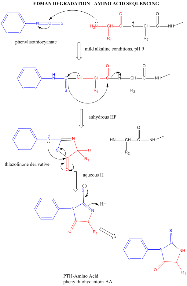
- Edman Degradation
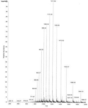
- ESI Mass Spectrum of Apo-Myoglobin
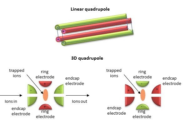
- Linear and 3D Quadrupoles
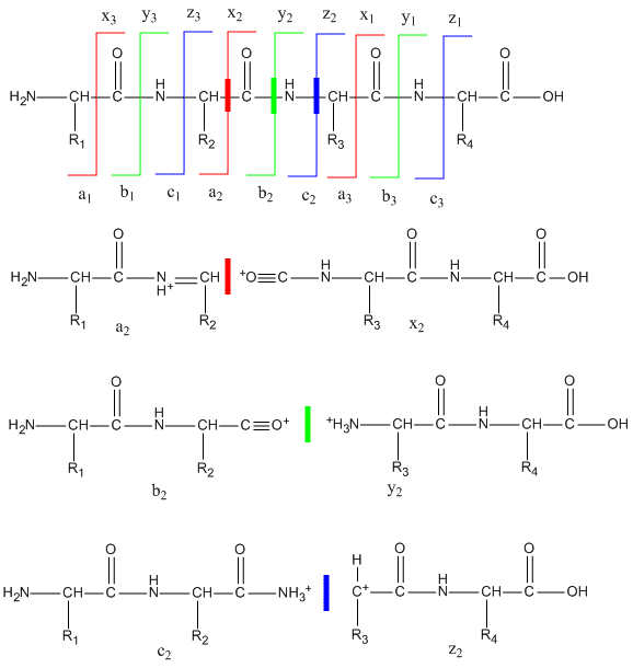
- Peptide Fragmentation and Sequencing by MS/MS
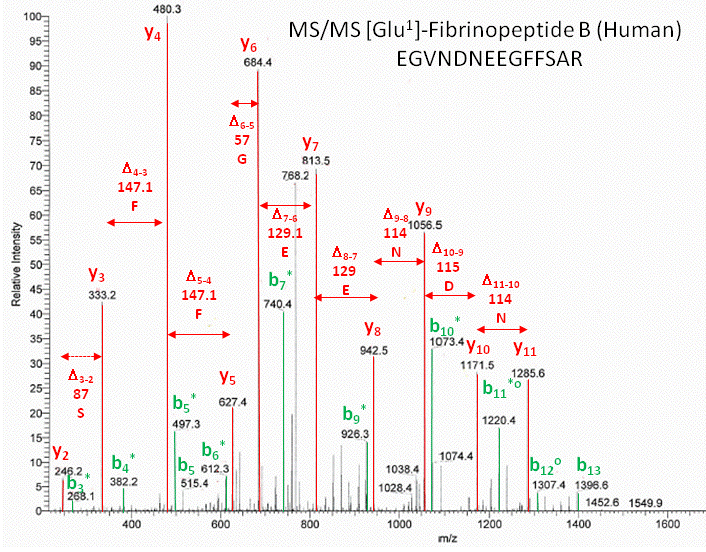
- Annotated MS/MS spectra of human Glu1- fibrinopeptide B
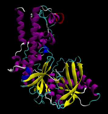
- Secondary structure
- Tertiary structure
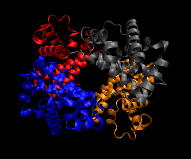
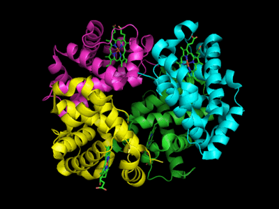
- Quaternary structure
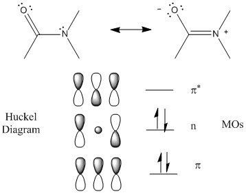
- Peptide Bonds - MOs and Huckel Diagram
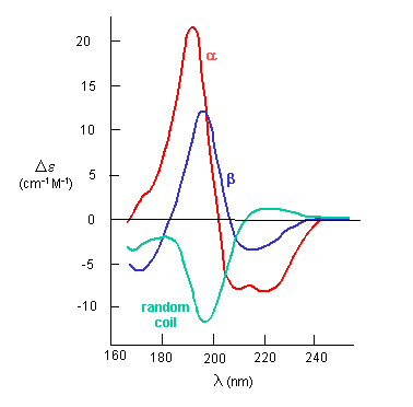
- The CD Spectra of Alpha-Helix, Beta-Sheet, and Random Coils
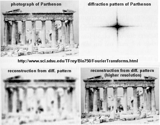
- X-Ray Structures and the Parthenon
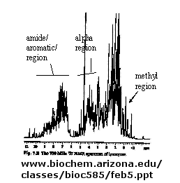
- 1D NMR spectra of a protein
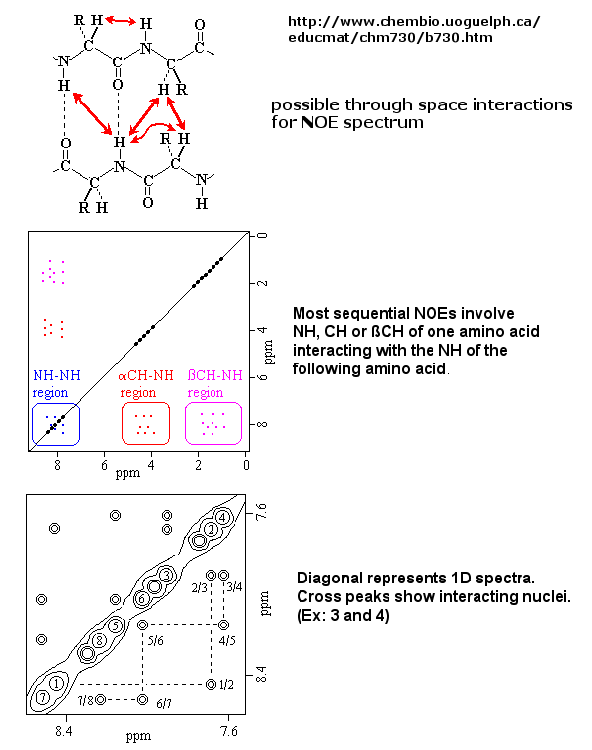
- 2D NOSEY spectra of a protein
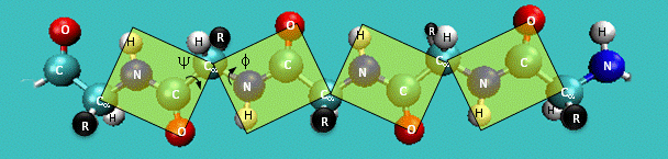
- Extended Polypeptide Showing Planes and Phi/Psi Angle
- Trans arrangement of the alpha C's
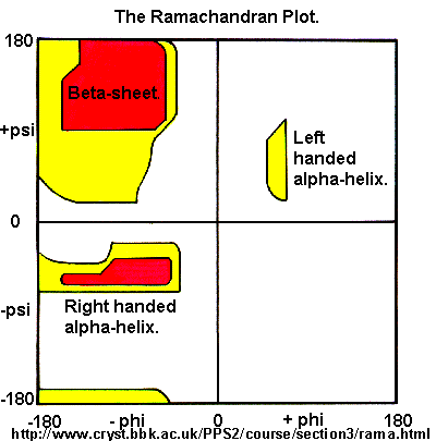
- Ramachandran Plot
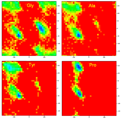
- Ramachandran plots showing Phi, Psi angles for Gly, Ala, Tyr, and Pro in actual proteins
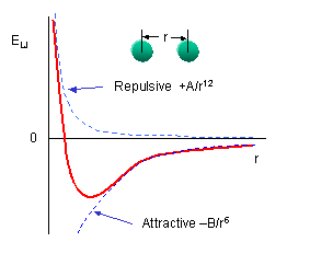
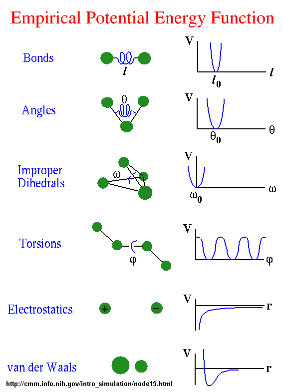
- Lennard-Jones potential (6-12 potential)
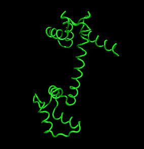
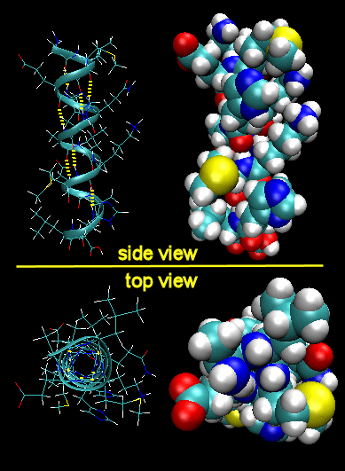
- Right Handed Alpha helices -
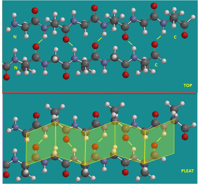
- Parallel beta strands
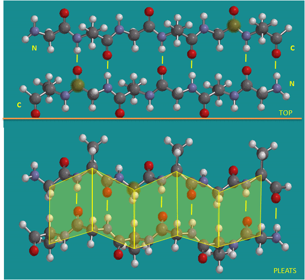
- Antiparallel beta strands
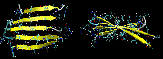
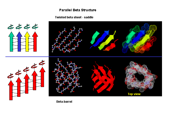
- Twisted Beta Sheet/Saddle
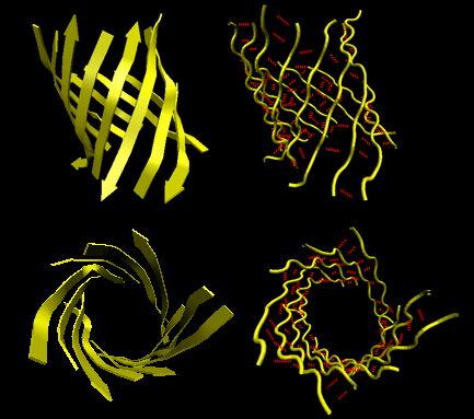
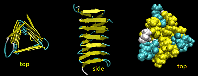
- Beta Barrel
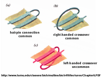
- Beta strand connections
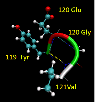
- Reverse Turns
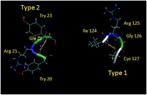
- Type I and Type II Reverse Turns
- Twisted sheets or saddles as well as beta barrels
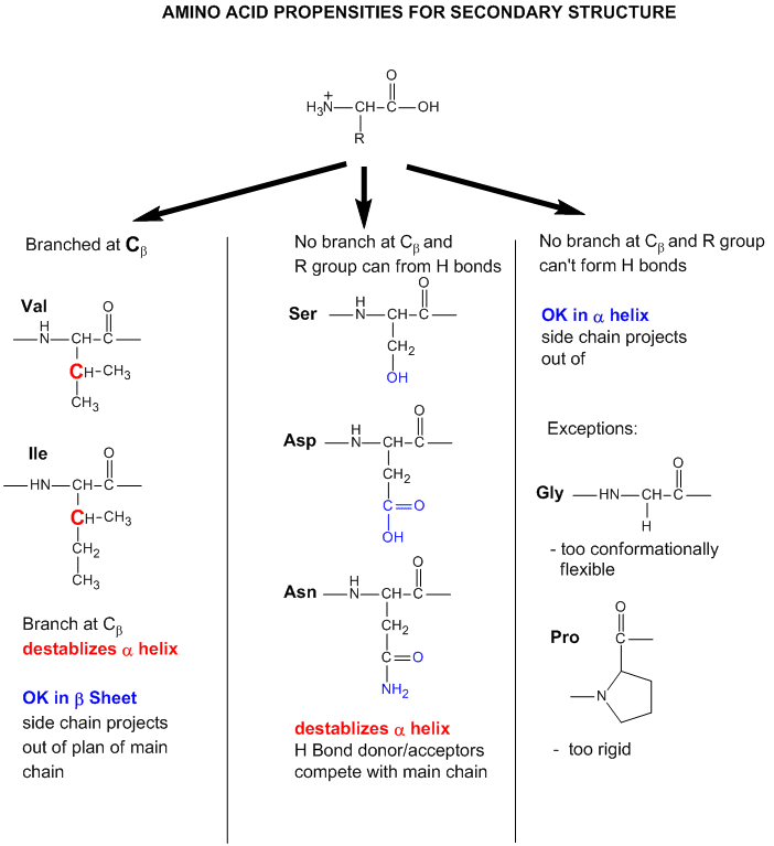
- Why do amino acid propensities for secondary structure differ?
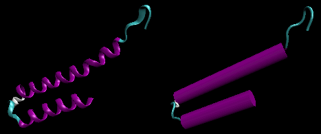
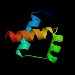
- Helix-loop-helix
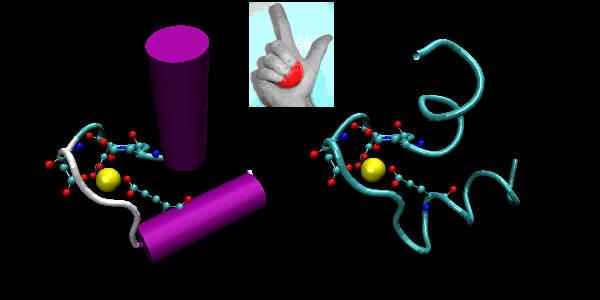
- EF hand.
- Beta-hairpin, or beta-beta
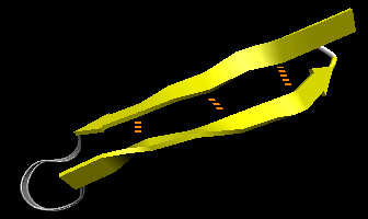
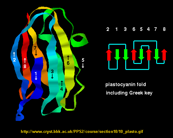
- Greek Key motif:
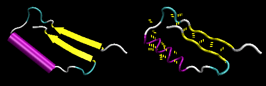
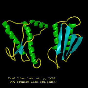
- Beta-alpha-beta:
- Beta Helices
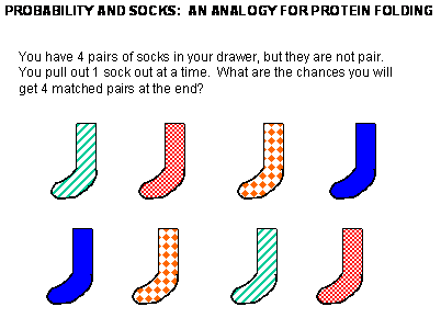
- Socks and protein folding
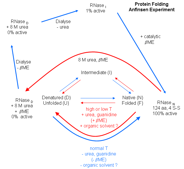
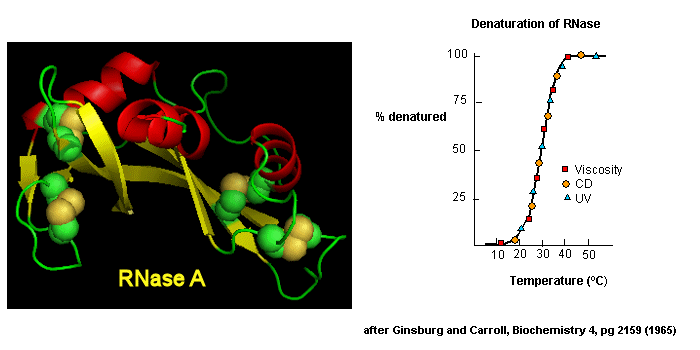
- Anfinsen Experiment: Folding of RNase
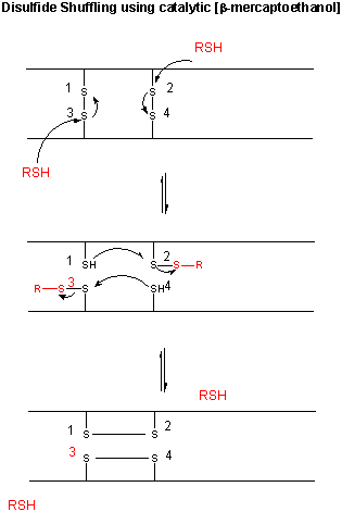
- Catalytic Shuffling of Disulfides with beta-mercaptoethanol
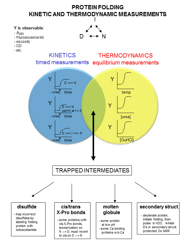
- Kinetic and thermodynamic measurements of proteins stability and folding
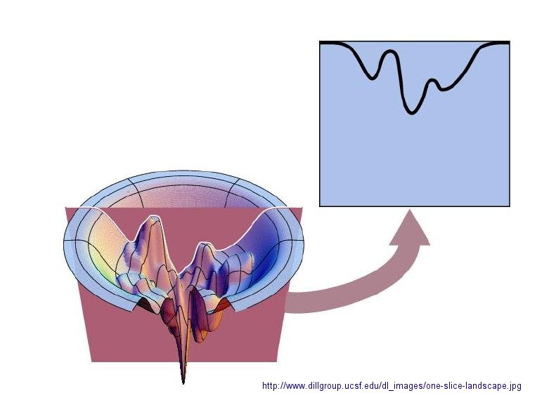
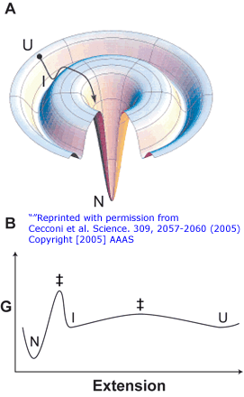
- Protein Folding Landscape:
- Reversible denatuation,
- Addition of iodoacetamide
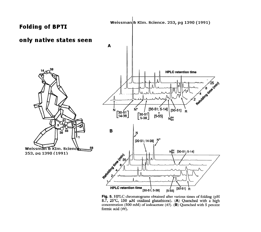
- Bovine pancreatic trypsin inhbitor (BPTI): Folding Kinetics
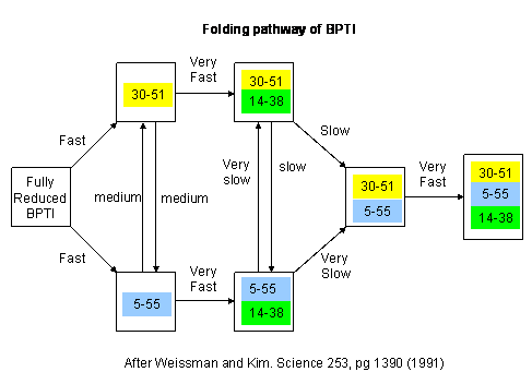
- BPTI: Folding Pathway In Vitro
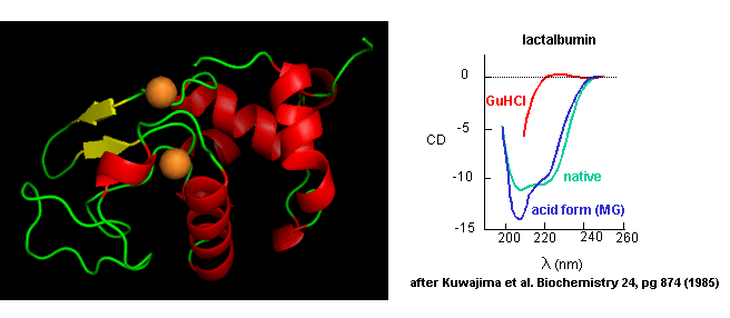
- Lactalbumin
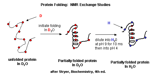
- NMR in protein folding: exchange of all C's with ionizable protons, including, the amide H's
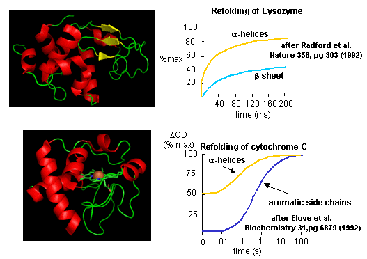
- Experimental data on model proteins
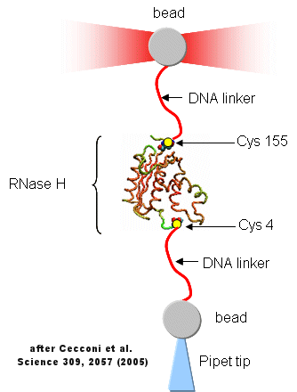
- Unfolding of RNase H with Optical Tweezers
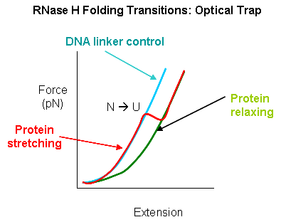
- RNaseH Folding Transitions: Optical Trap
- Folding Lanscape of RNase H: Optical Tweezers
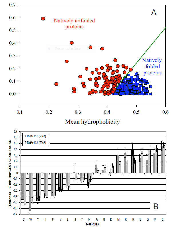
- Characteristics of Intrinsically Disordered Proteins
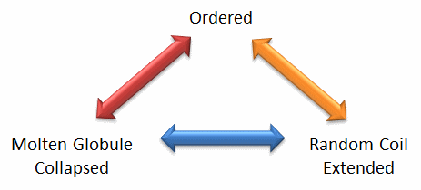
- The Protein Trinity: Ordered, Collapsed and Extended States
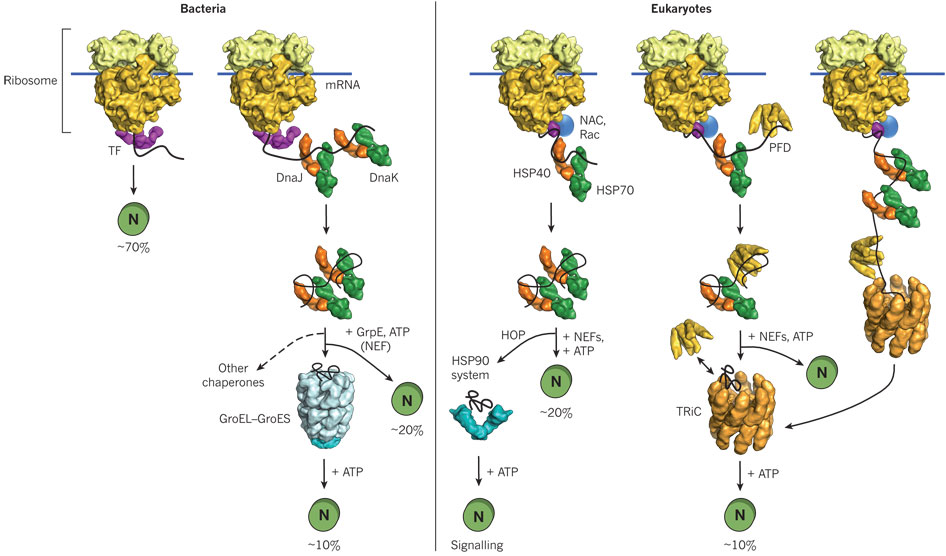
- Comparison of cytosolic prokaryotic and eukaryotic chaperone pathways
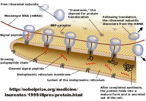
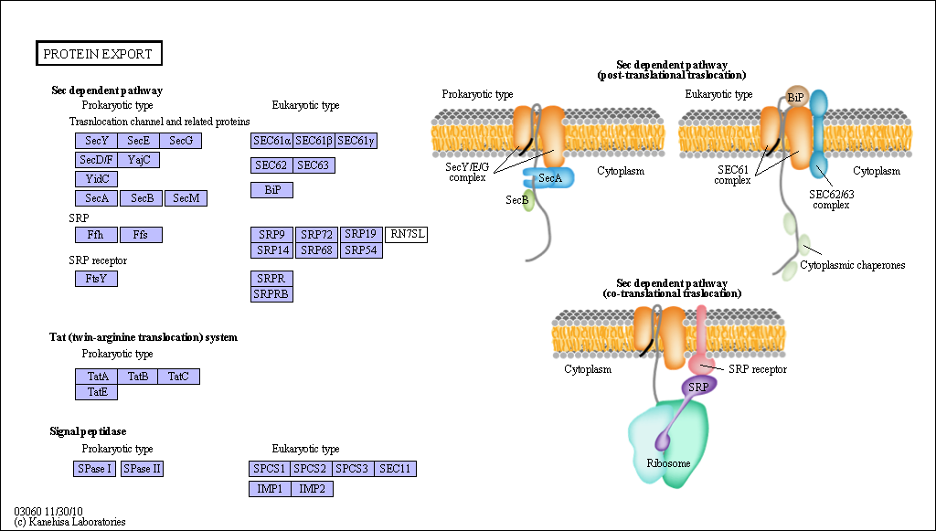
- Overview - Synthesis of Protein Destined for Secretion
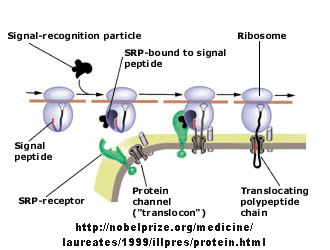
- Signal Recognition Particle Complex
- Sec Dependent Pathway for Post-translational translocation
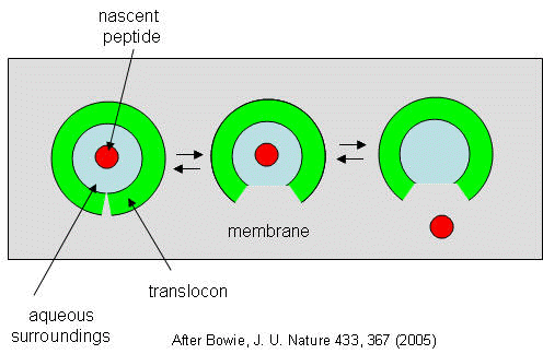
- Translocon Equilbrium Model
- GroEL and GroES.
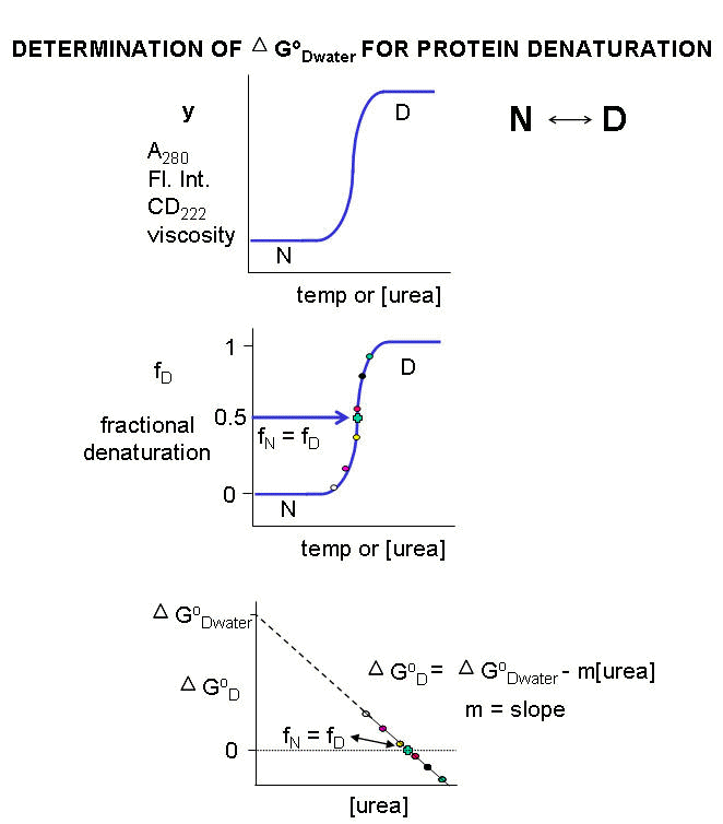
- Denaturation Curves for Proteins
- Diagram showing relative contributions to the ΔG for protein folding.
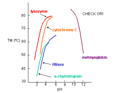
- Denaturation temperature vs pH for proteins
- Thermodynamic cycle for formation of a H-bonded dimer of N-methylacetamide in a nonpolar solvent
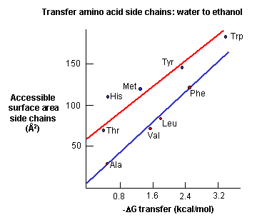
- Free energy of transfer of amino acids from water
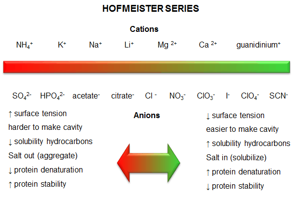
- Hofmeister Series
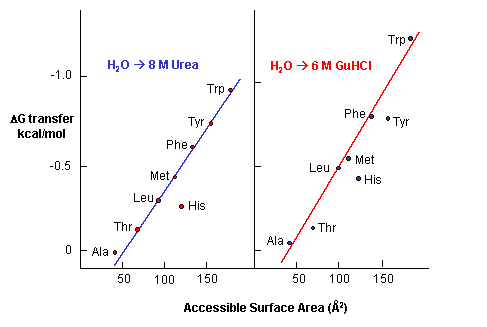
- Free energy of transfer of the nonpolar amino acids into 8M urea
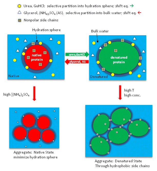
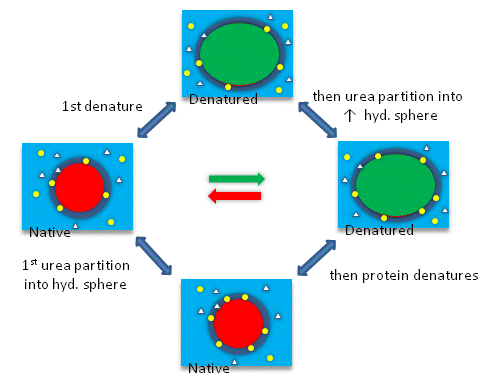
- Reagents might interact with the surface of the protein.
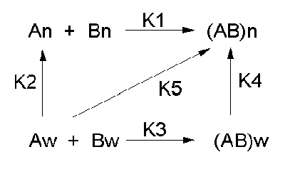
- Thermodynamic cycle for urea denaturation of proteins
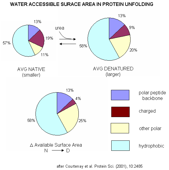
- Water Accessible Surface Area in Protein Unfolding
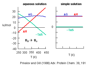
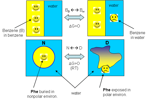
- Change in G, H and -TS for taking benzene from pure benzene to water.
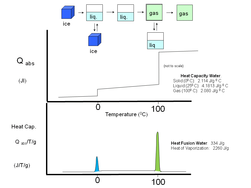
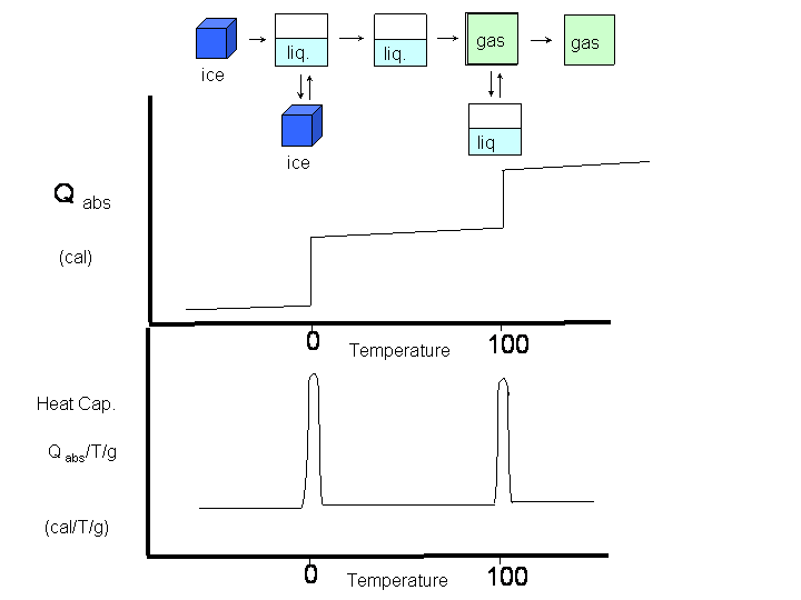
- Heat of fusion, heat of vaporization, and heat capacity of water
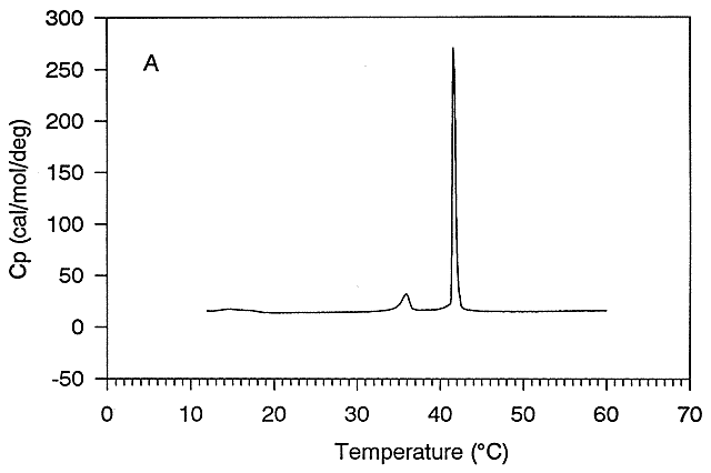
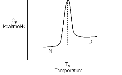
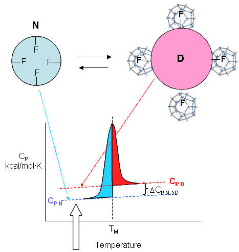
- Thermal Denaturation of a protein using differential scanning calorimetry
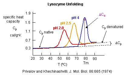
- Heat capacity of proteins vs temperature
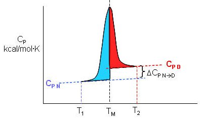
- Determination of ΔH for Protein Denaturation by Differential Scanning Calorimetry
- Heat Capacity Changes: A Molecular Interpretation
- Analogy between benzene solubility in water and protein denaturation
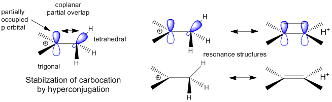
- Stabilization of Carbocations by Hyperconjugation
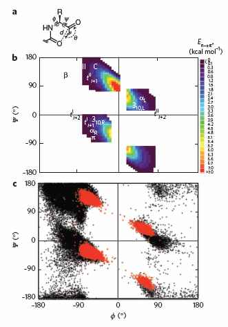
- Orbital involvement in H-bonds and n to pi* interactions in alpha helices
- Ramachandran plots of n to pi* interactions.
- Amino Acid Structure and propensity for secondary structure
- Secondary structure prediction
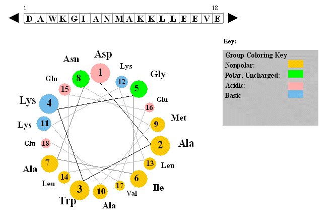
- Helical wheel projection
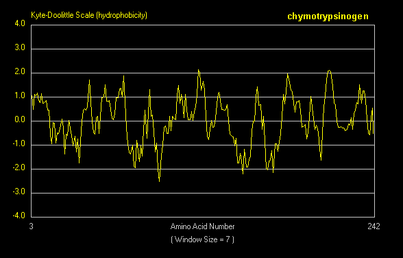
- Hydrophathy plot for chymotrypsinogen
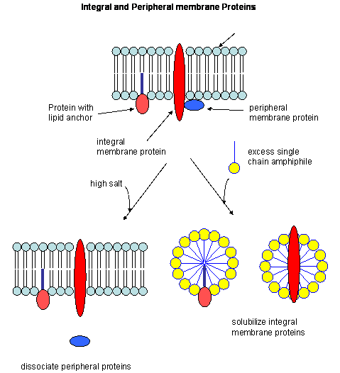
- Types of membrane proteins
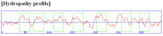
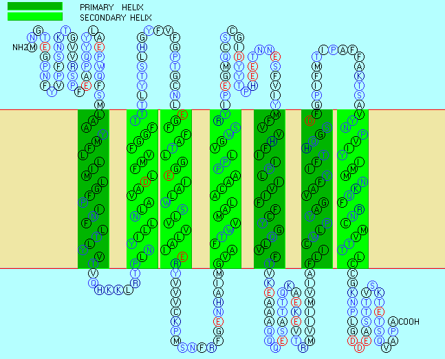
- Hydropathy plot for bovine rhodopsin
- Seven transmembrane helices for bovine rhodopsin
- Nanodisc with membrane protein
- 3-helix bundle
- Helices form early
- Simultaneous and quick partial helix formation and collapse
- Engrailed homeodomain 3 helix bundle protein
- Complete Folding Pathway of Engrailed Homeodomain by Experiment and Simulation
- Alzheimer's protein: beta amyloid protein and mutations
- Cartoon Models of PrPc and PrPsc
- Potential energy functions in protein mechanics and dynamics calculations
Chapter 3: Carbohydrates
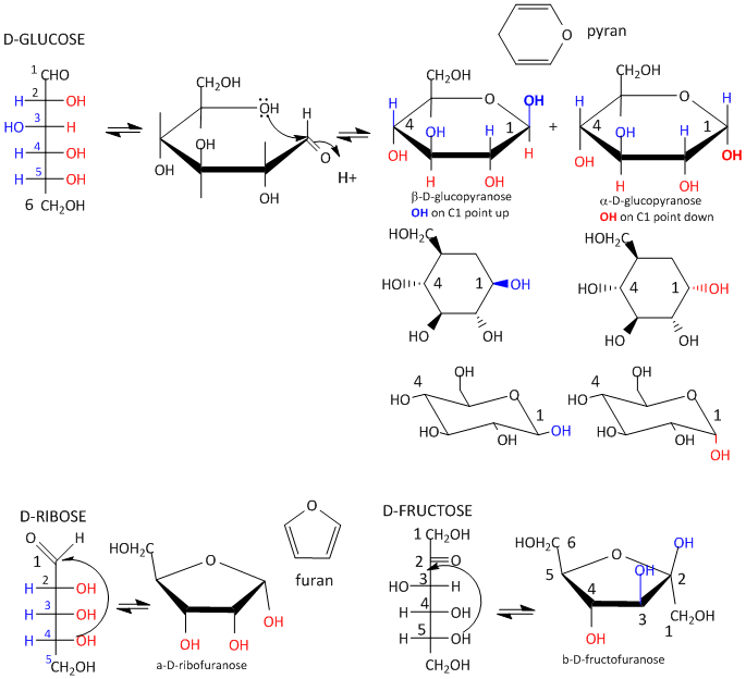
- Glucose/fructose cyclization
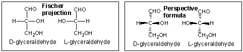
- Glyceraldehyde perspective structures
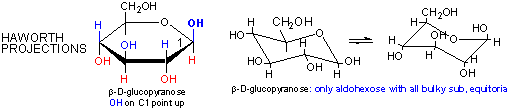
- Glucose: Haworth and chair structures
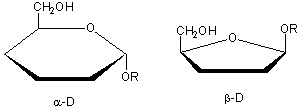
- Alpha/beta anomers
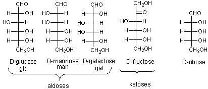
- Monosaccharides to know
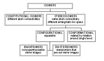
- Isomer review
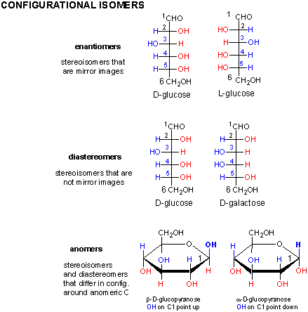
- Sugars: configurations isomers
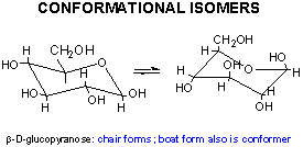
- Sugars: conformational isomers
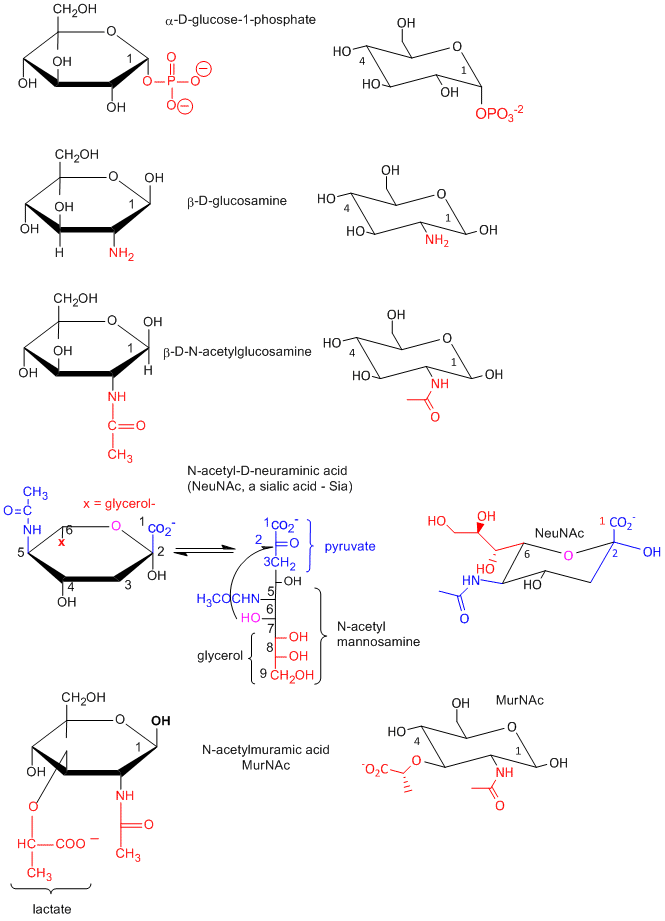
- Sugar derivatives
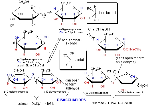
- Hemiacetal/acetal chemistry
- Dissacharides: lactose and sucrose
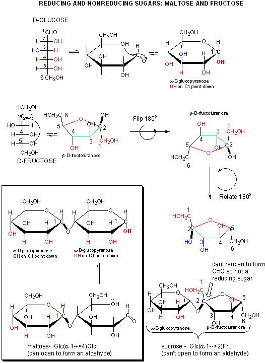
- A closer look at reducing and nonreducing sugars: lactose and maltose
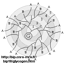
- Glycogen
- Homopolysaccharides in Chair Conformations
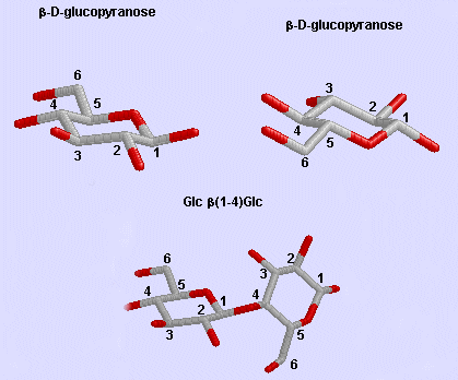
- Glc β (1-4) Glc link
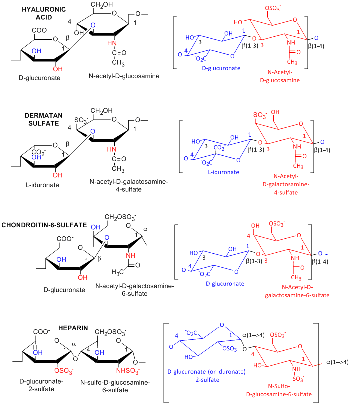
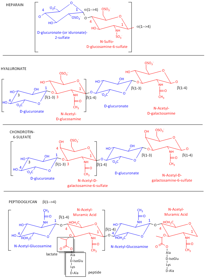
- Glycosaminoglycans
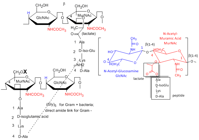
- Peptidoglycans
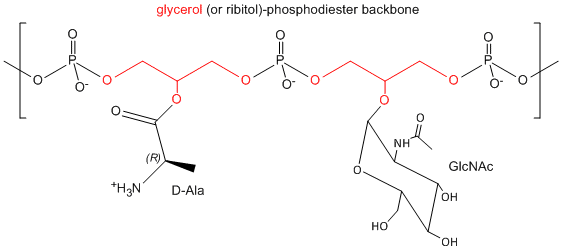
- Teiochoicacid
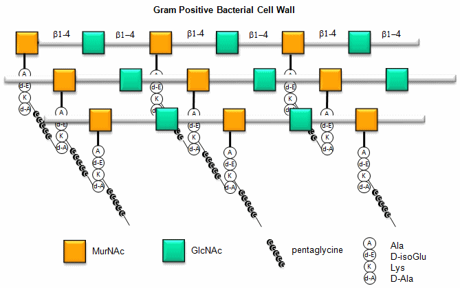
- Gram positive membrane structure (after)
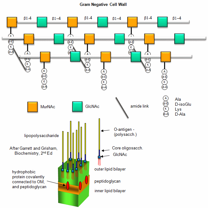
- Gram negative membrane structure (after)
- Heteropolysaccharides in Chair Conformations
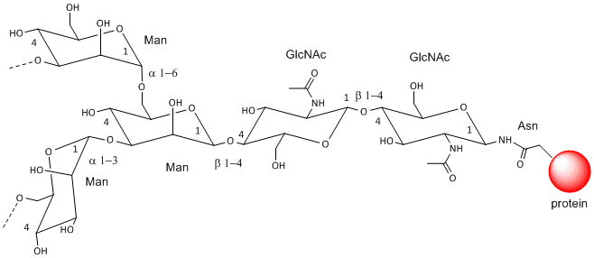
- N-linked glycoproteins
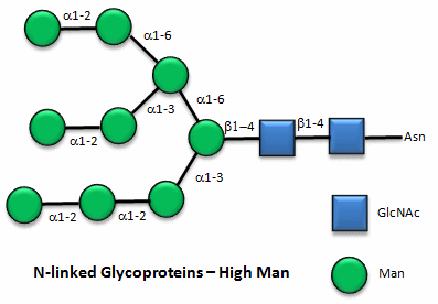
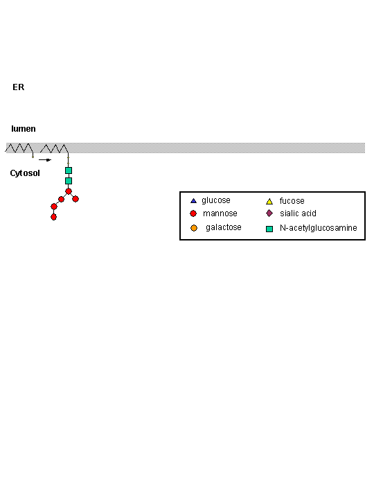
- N-linked high mannose glycoproteins
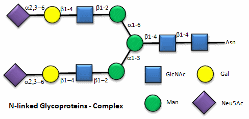
- N-linked complex glycoproteins
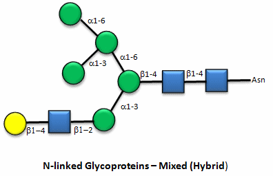
- N-linked hybrid glycoproteins
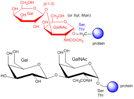
- O-linked Glycoproteins
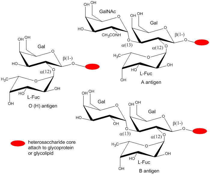
- Blood Group Antigens
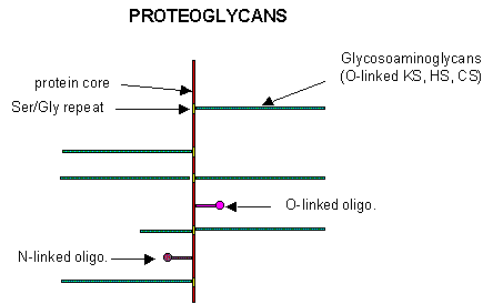
- Proteoglycans
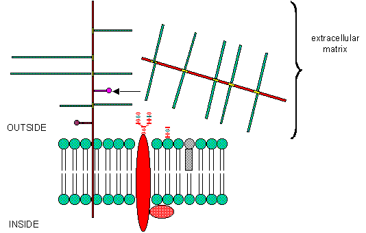
- Extracellmatrix
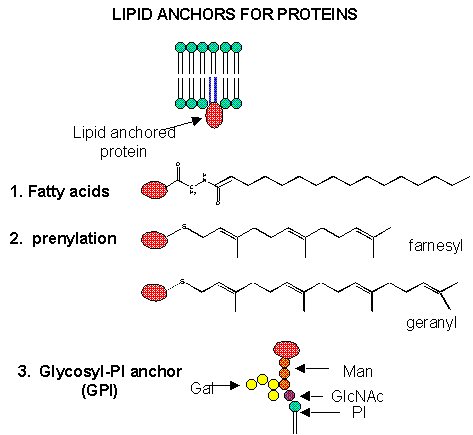
- Lipid anchors
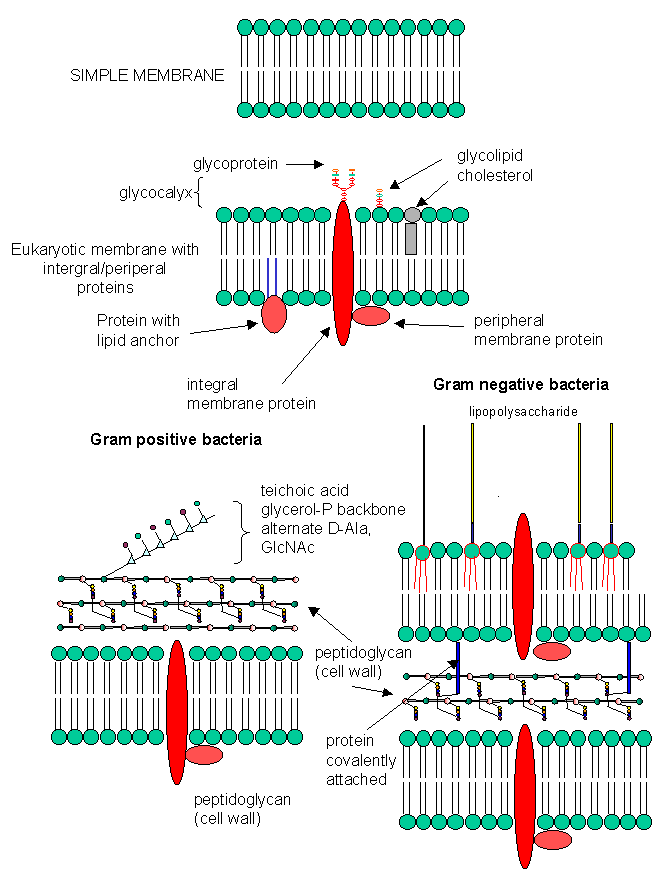
- Biological Membranes: Simple to Complex
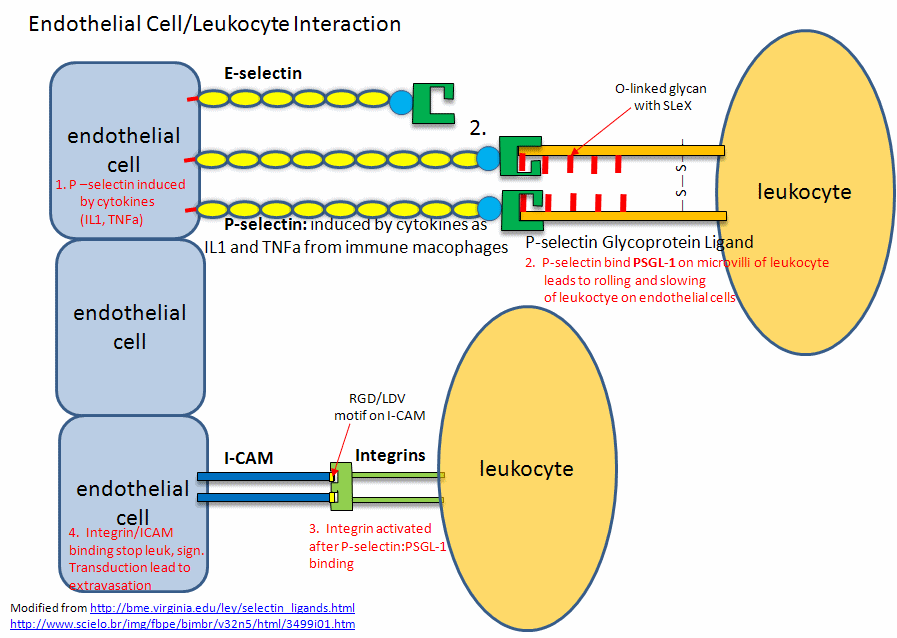
- Endothelial Cell/Leukocyte Interactions: Selectins, Integrins, and ICAMs
- A cool view of a membrane surface
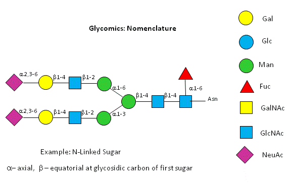
- CHO symbolic nomenclature
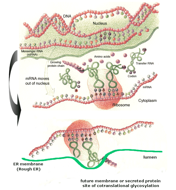
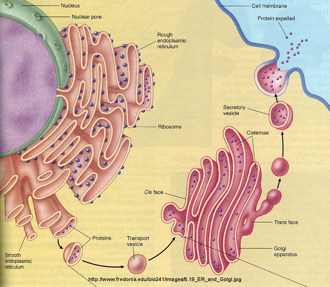
- Ribosomes bind to an elongated, extensive organelle in the cell called the endoplasmic reticulum (ER).
- Endoplasmic reticulum
- Golgi apparatus
- Golgi
- Calnexin pathway
Chapter 4: DNA, Genomics, and Proteomics
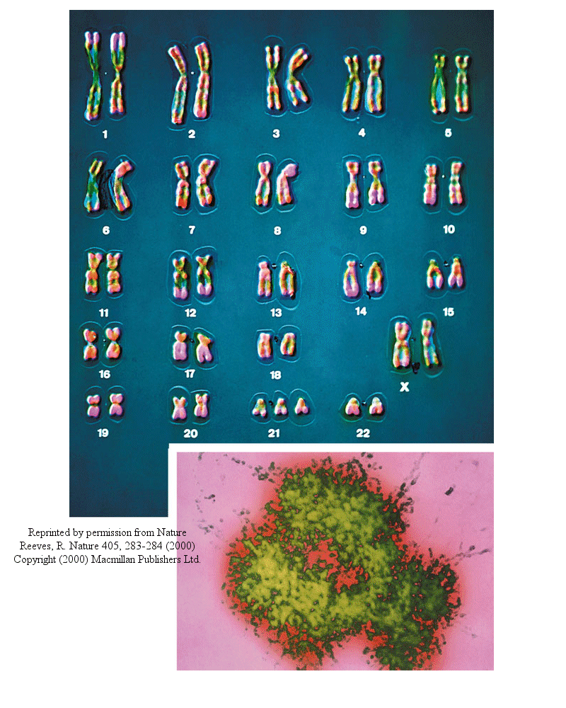
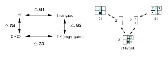
- Human Chromosomes (with an extra copy of Chromosome 21, which causes Down syndrome)
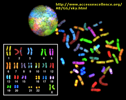
- Spectralkaryotype chromosome analysis
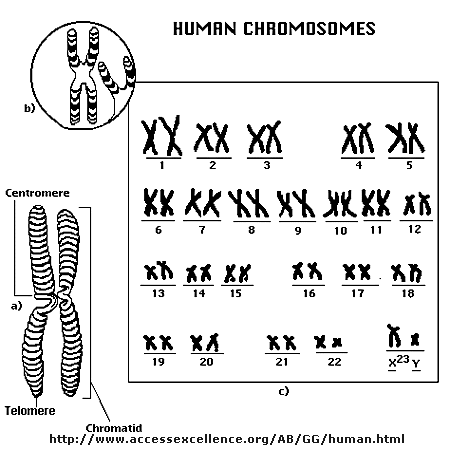
- Human Chromosomes
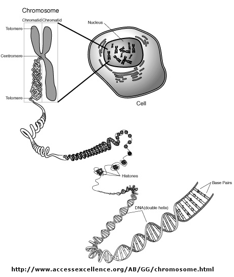
- Packaging of DNA in the Nucleus
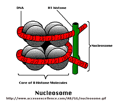
- Nucleosome
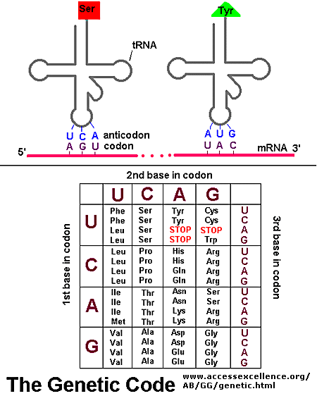
- Codon:Anticodon interactions between mRNA and tRNA
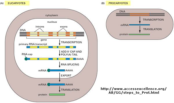
- Central Dogma Differences in Eukaryotes and Prokaryotes
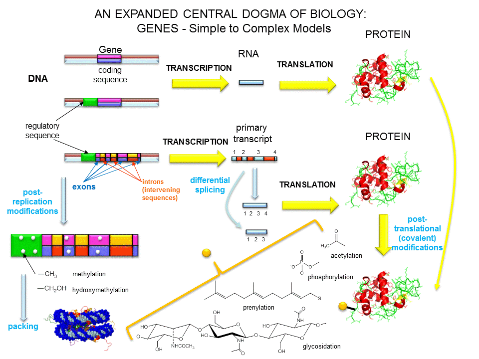
- A view of genes and their products: simplicity to Complexity
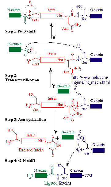
- Mechanism of Intein Splicing
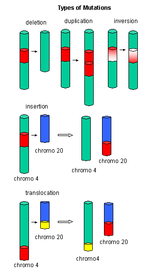
- Mutations
- Another View of Mutations
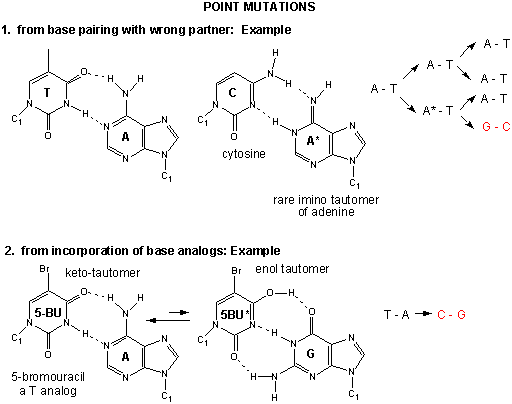
- Point mutations from mismatch pairing and Incorporation of Base Analogs
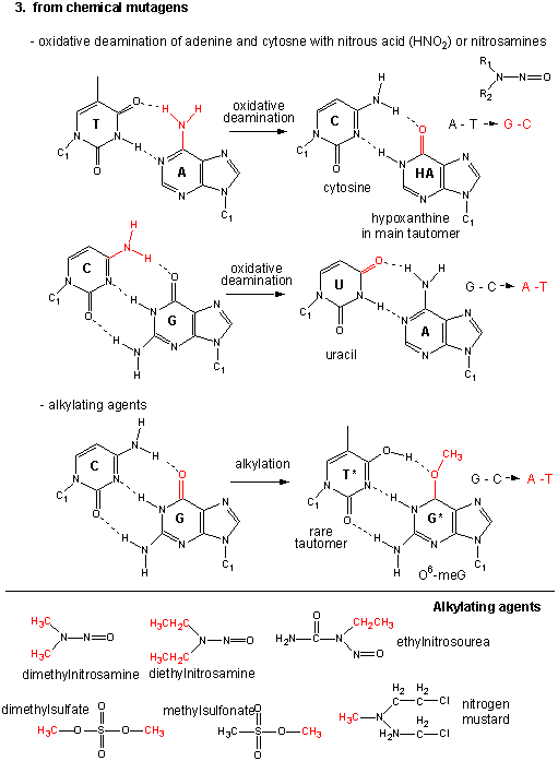
- Point Mutations from alkylating Agents
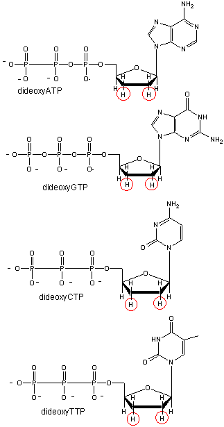
- Didexoynucleotides
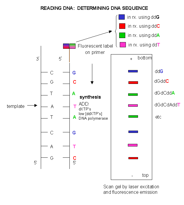
- DNA sequencing using different fluorescent primers for each ddXTP reaction
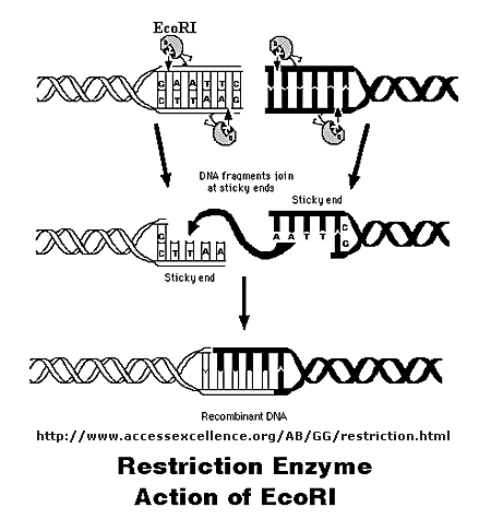
- Cleaving DNA with the Restriction Enzyme EcoR1
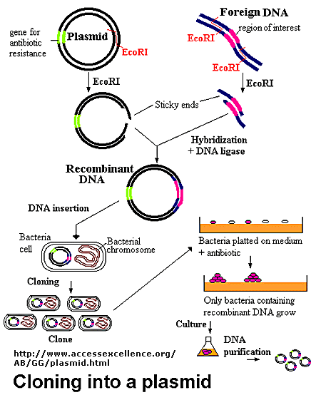
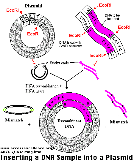
- Cloning a Restriction Fragment into a Plasmid
- Another View
- Introns and Exons: Central Dogma in Eukaryotes and Prokaryotes
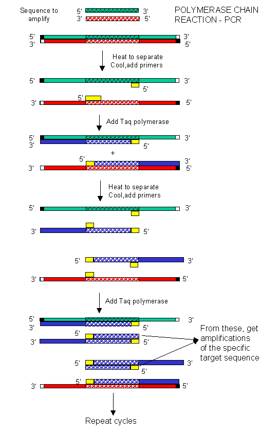
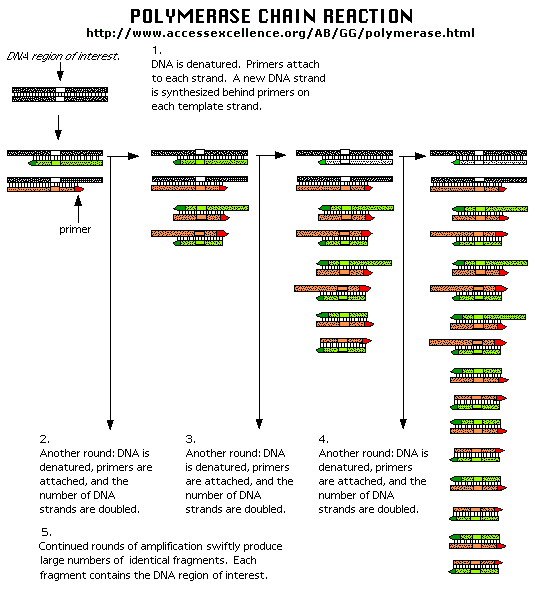
- Copying DNA in the test tube - the polymerase chain reaction (PCR)
- Another View of PCR
- Site Specific Mutagenasis
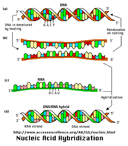
- Nucleic Acid Hybridization
- Finding a mRNA sequence of minimal degeneracy.
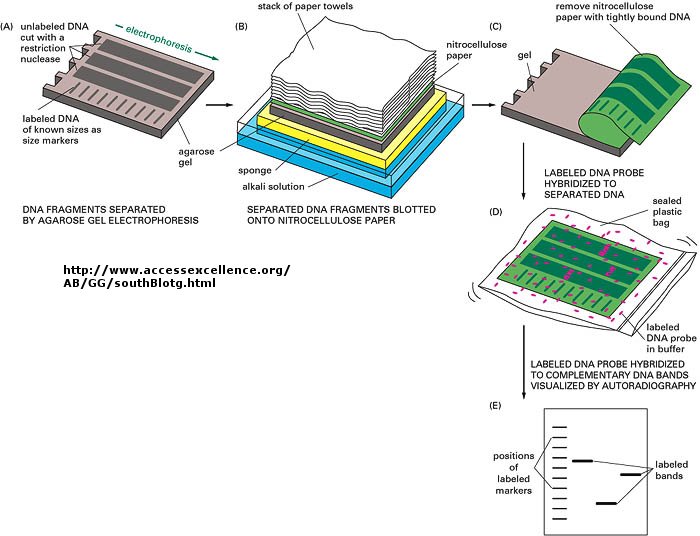
- Southern Blotting
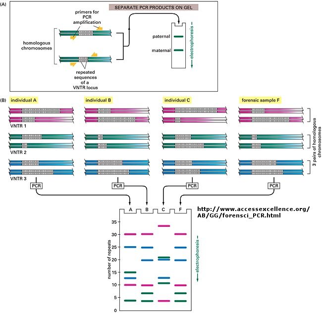
- Southern blot/PCR forensic analysis - restriction fragment length polymorphism (RFLP)
Chapter 5: Binding
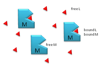
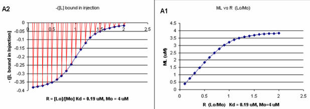
- M is free macromolecule, L is free ligand, and ML is macromolecule-ligand complex,
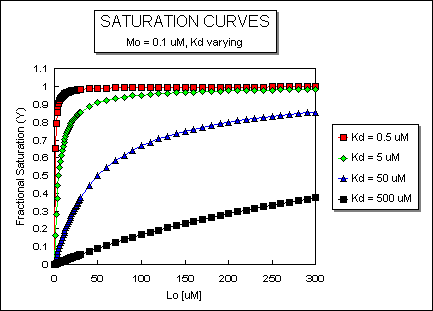
- ML vs L at Varying Kd's
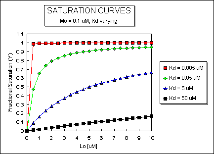
- ML vs L at Even Lower Kd's
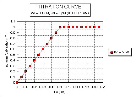
- ML vs L at a Very Low Kd!
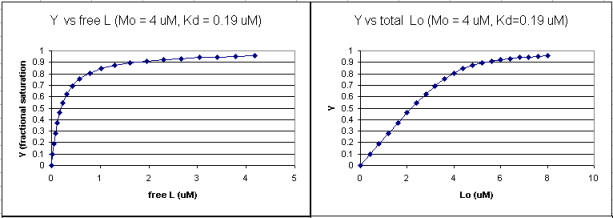
- Y vs L and Y vs Lo when Lo is not >> Mo
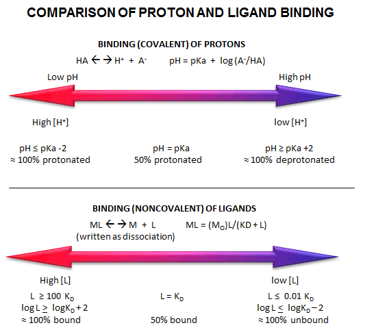
- Comparison of Covalent Binding of Protons vs Noncovalent Binding of Ligand
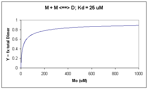
- Saturation binding curve for dimerization of a macromolecule
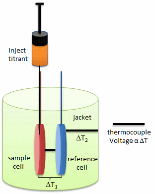
- Isothermal Titration Calorimeter Cells
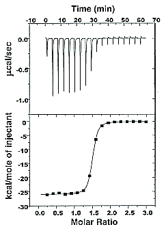
- Typical isothermal titration calorimetry data and analysis
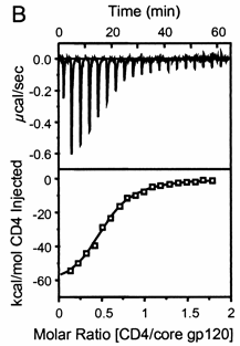
- Titration calorimetry determination of Kd and ΔH for the interaction of gp120 and CD4
- Binding Curves that Explain Sigmoidal Titration Calorimetry Data for gp120 and CD4
- Surface Plasmon Resonance
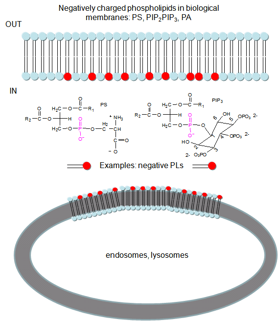
- Negatively charged phospholipids in biological membranes
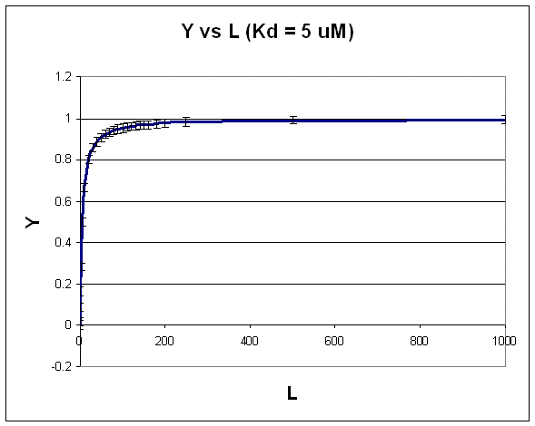
- Hyperbolic plot: Y vs L
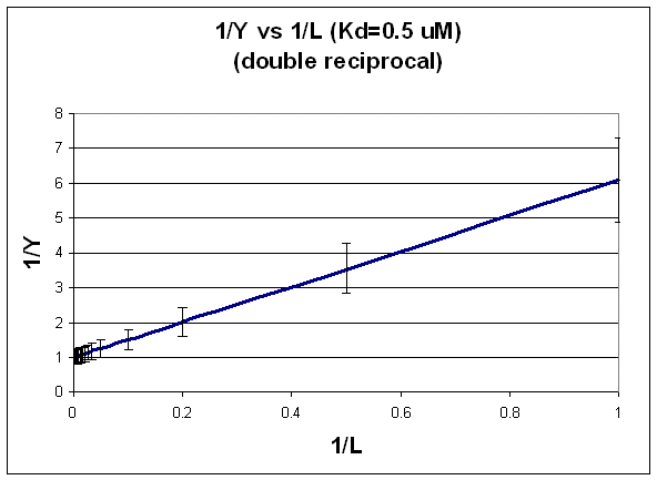
- Double-reciprocal plot: 1/Y vs 1/L
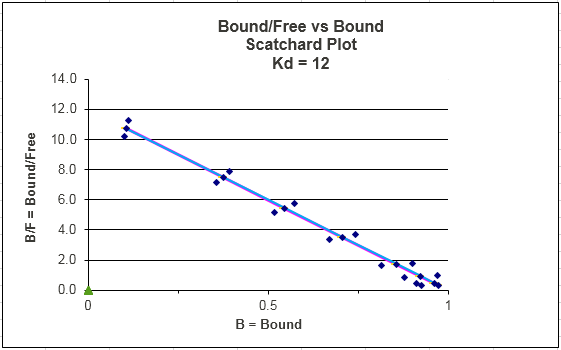
- Scatchard plot: Y/L vs Y
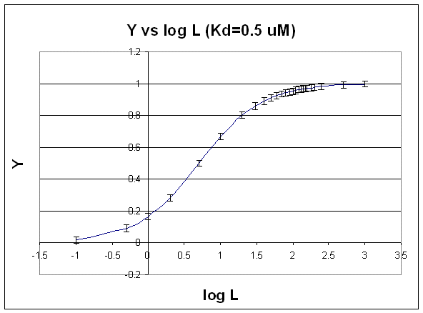
- Semilog plot: Y vs log L
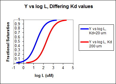
- Y vs log L: Differing Kd values
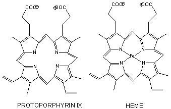
- Heme
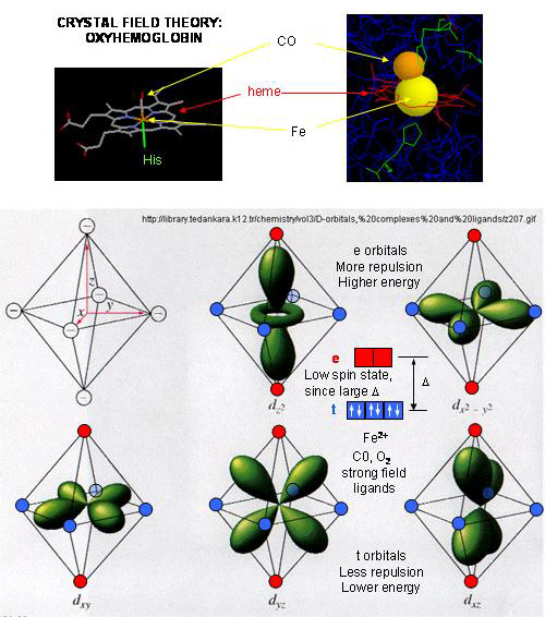
- Heme-O2 Octahedral Complex
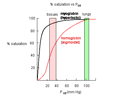
- Plots of Y (fractional saturation) vs L (pO2) are hyperbolic for Mb, but sigmoidal for Hb,
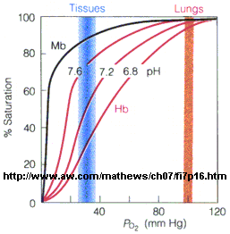
- Bohr Effect - Hemoglobin
- Conformation Changes on Oxygen Binding to Deoxy-Hemoglobin
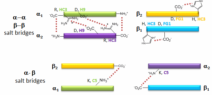
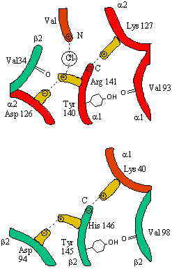
- Salt Bridges in Deoxy Hb
- Salt Bridges in DeoxyHb
- C- or N- terminus
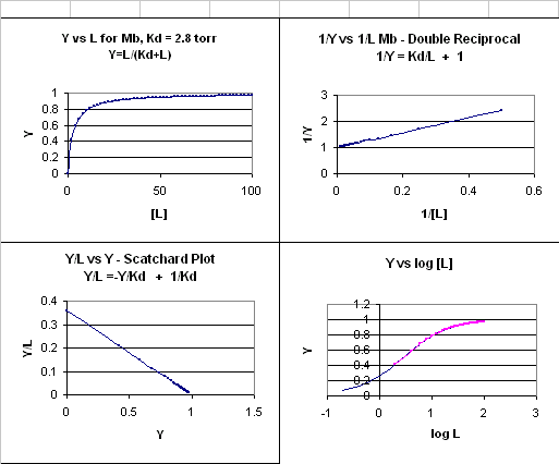
- 4 Ways to plot Mb and O2 plots
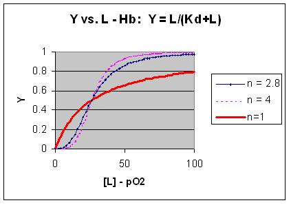
- Plots of Y vs L for Hb with varying degrees of cooperativity: n = 1, 2.8, and 4
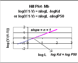
- Hill Plot for Mb (n =1)
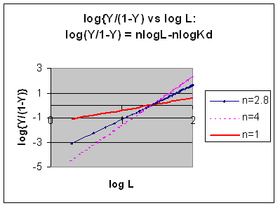
- Hill Plots: n = 1, 2.8, and 4
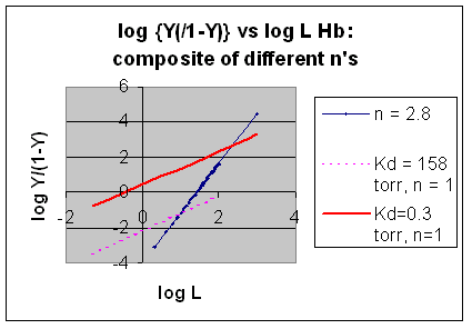
- Hill Plots For Hb Showing straight lines for n=2.8 and for n's=1 which model the low and high affinity sites.
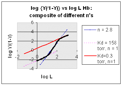
- Hill Plot for Hb: Black line showing hypothetical actual curve
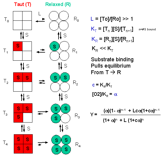
- Linked equilibrium in he MWC model
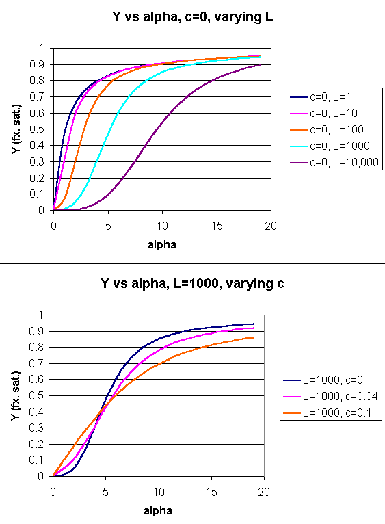
- Y Vs alpha with varying L and c - MWC MODEL
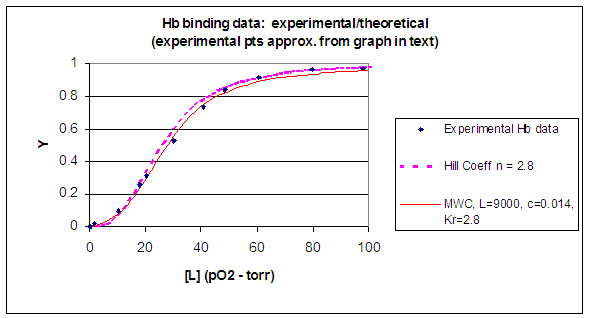
- Hb binding curves: Experimental, vs Theoretical Hill and MWC Equations
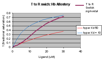
- Cooperativity as transition between two hyperbolic binding curves with different Kd's
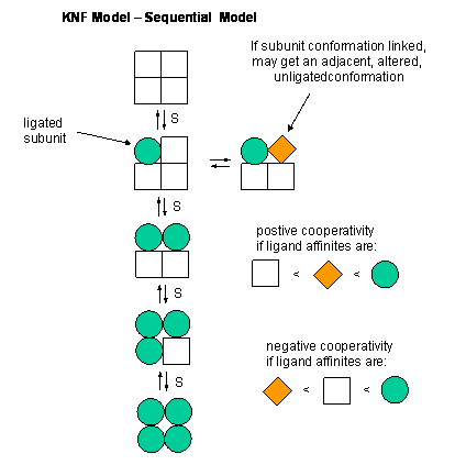
- Equilbria in KNF Model
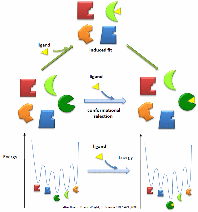
- Conformational Selection vs Induced Fit Binding
-
Types of Molecular Recognition Features in Intrinsically Disordered
Proteins
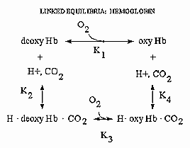
- Linked equilibrium: binding of H+, CO2 and O2 to hemoglobin
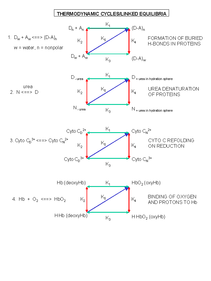
- A review of thermodynamic cycles - linked equilibria
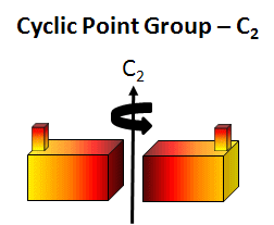
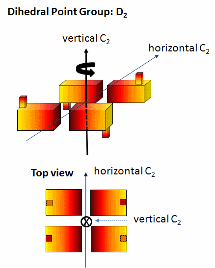
- C2 Symmetry
- D2 Symmetry
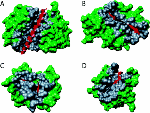
- Mutations causing Dimer with C2 symmetry or Infinite Helix
- Equilibria in KNF Model
- Conformational Selection vs Induced Fit Binding
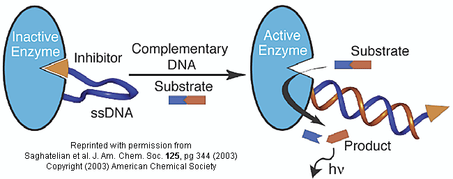
- Inhibitor-DNA-enzyme (IDE) - an engineered allosteric enzyme
- A combination of MWC and KNF: a modern structure view of structural changes in Hb on ligand binding
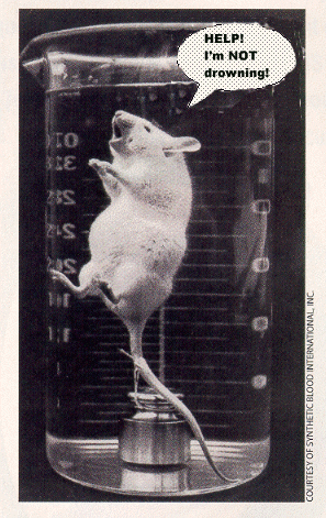
- Of mice and oxygen
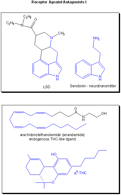
- Receptor agonists/antagonist I
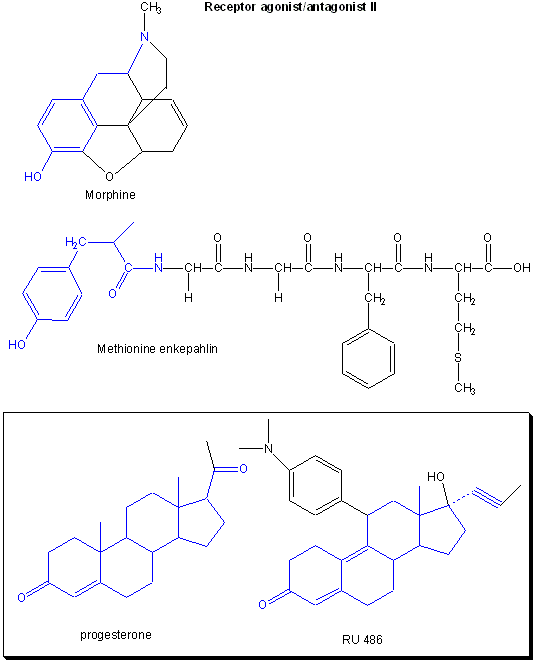
- Receptor agonists/antagonist II
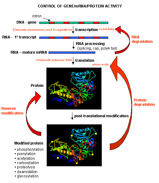
- Process that affect the steady state concentration of a protein
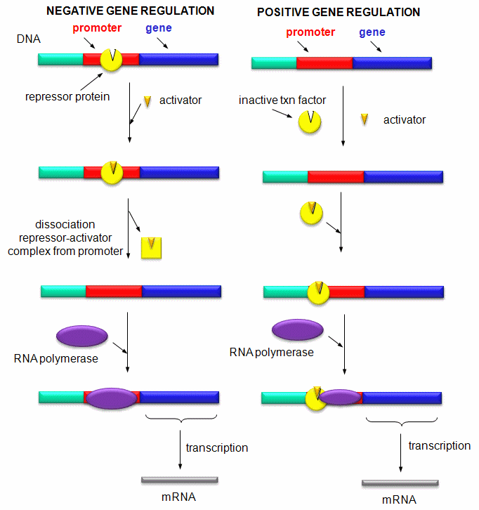
- Positive and Negative Regulation of Gene Transcription
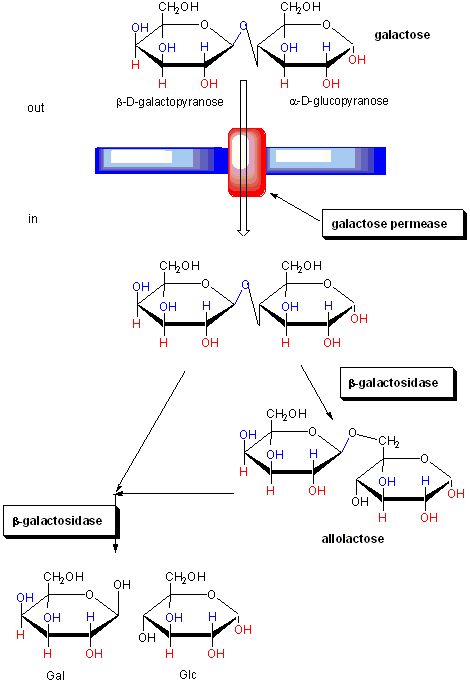
- Function of proteins in galactose utilization
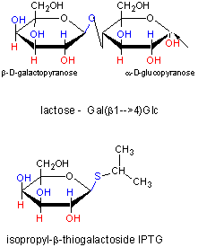
- IPTG and Lactose Structures
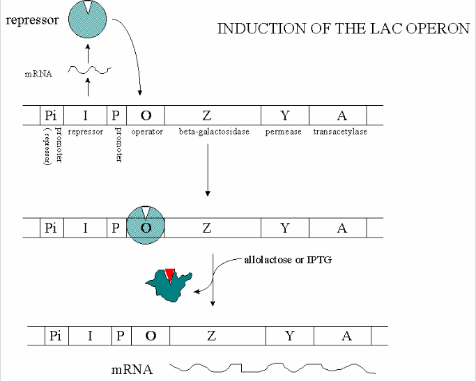
- Induction of the Lac Operon
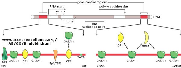
- Control of globin gene transcription
- Example of transcription complexes
- Upon absorbing red light, a phytochrome photoreceptor is converted from the inactive Pr form to the active Pfr form (permission)
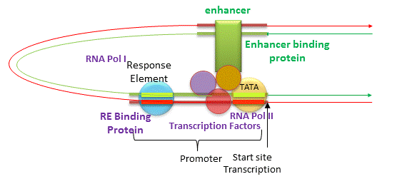
- Enhancers of Transcription
- Eukaryotic promoters and regulatory regions
- Eukaryotic multisubunit general transcription apparatus
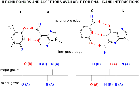
- AT and GC base pairs have available H bond donors and acceptors
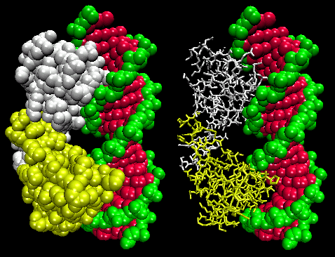
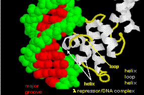
- Lambda Repressor/DNA Complex
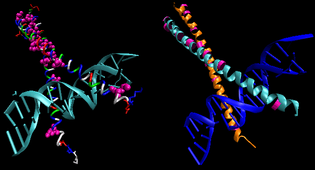
- helix-turn-helix in λrepressor/DNA complex
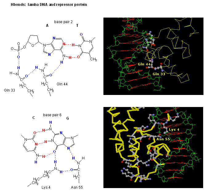
- H Bond interactions betweenλ repressor and DNA
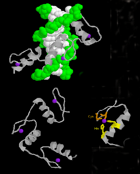
- Zinc finger
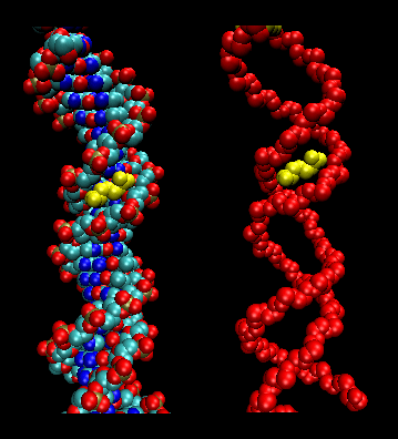
- Leucine zippers
- Arg in T3c Transposase binding in Narrowed Minor Grove of T3c Transposon
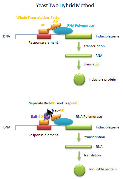
- Yeast Two-Hybrid
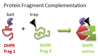
- Protein Fragment Complementation
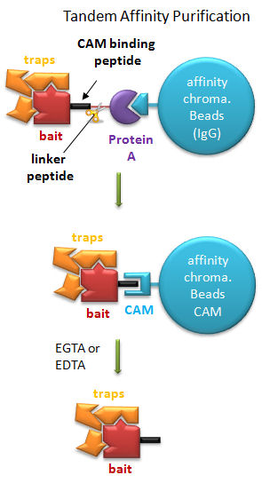
- Tandem Affinity Purification
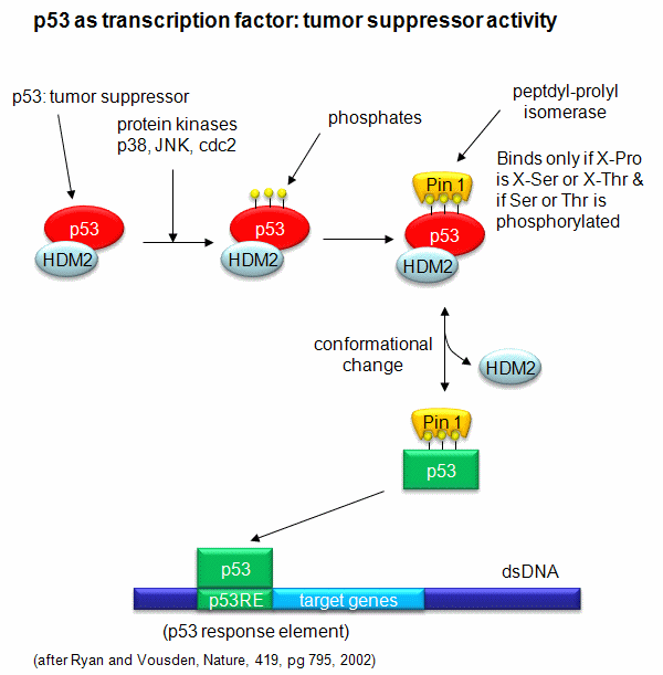
- Activation of p53 as a transcription factor by phosphorylation and conformational change
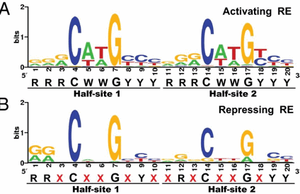
- Canonical sequence for both activating and repressing p53RE
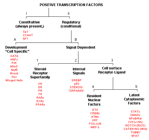
- Transcription Factors: Functional Classification
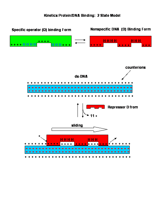
- Kinetics of protein-DNA binding
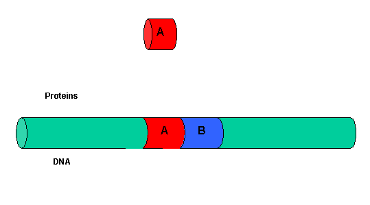
- Two proteins, A and B,
- Cooperative binding of B to the protein A-DNA complex.
- Chromatin
- Nucleosome
- Remodeling of Chromatin and Control of DNA Transcription
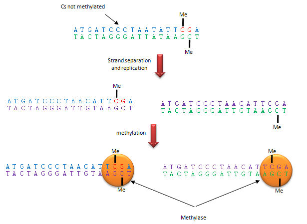
- Methylation of CpG
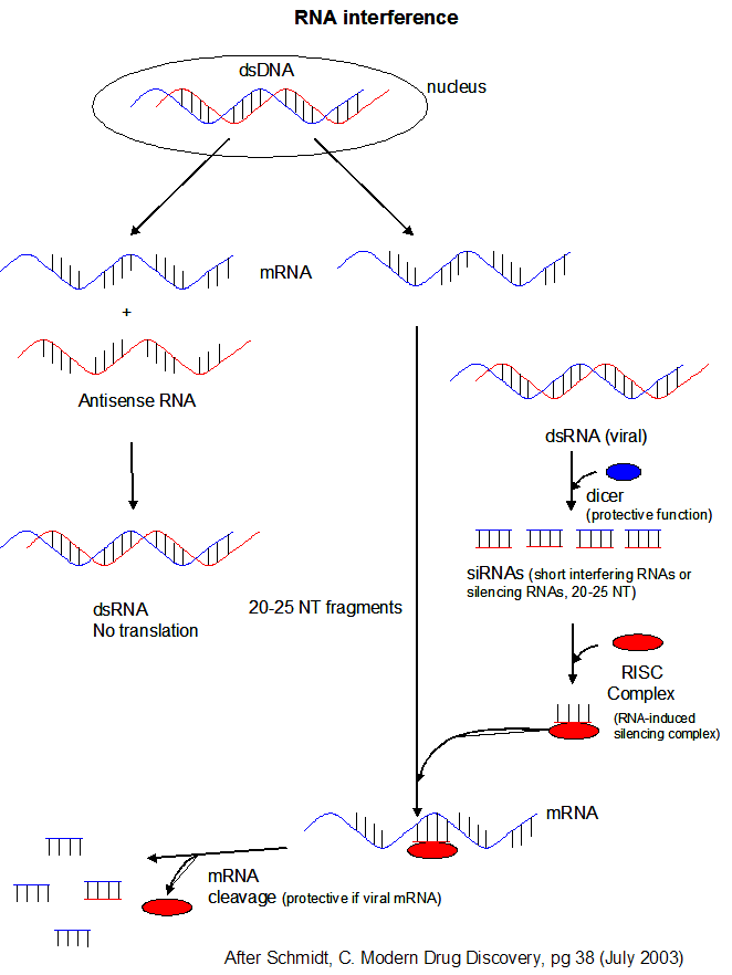
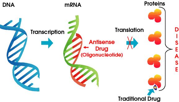
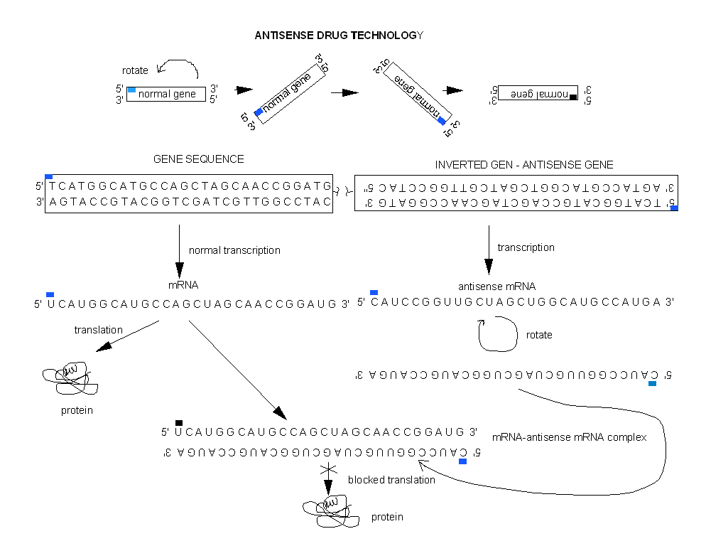
- RNA Interference: Antisense and Silencing
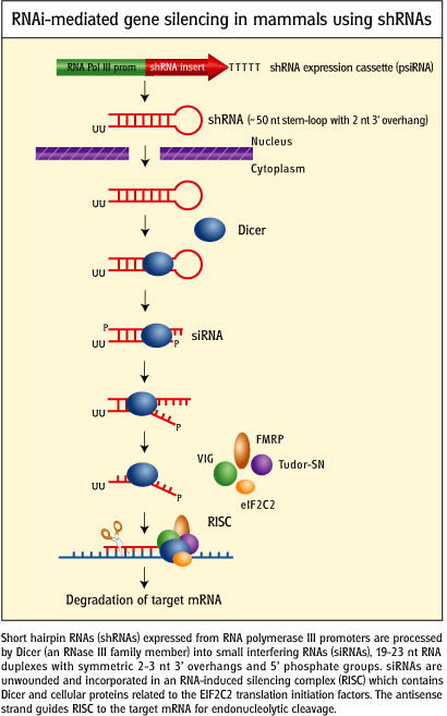
- RNAi-mediated gene silencing in mammals using short haripin RNA genes.
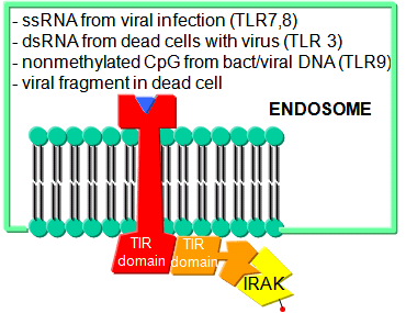
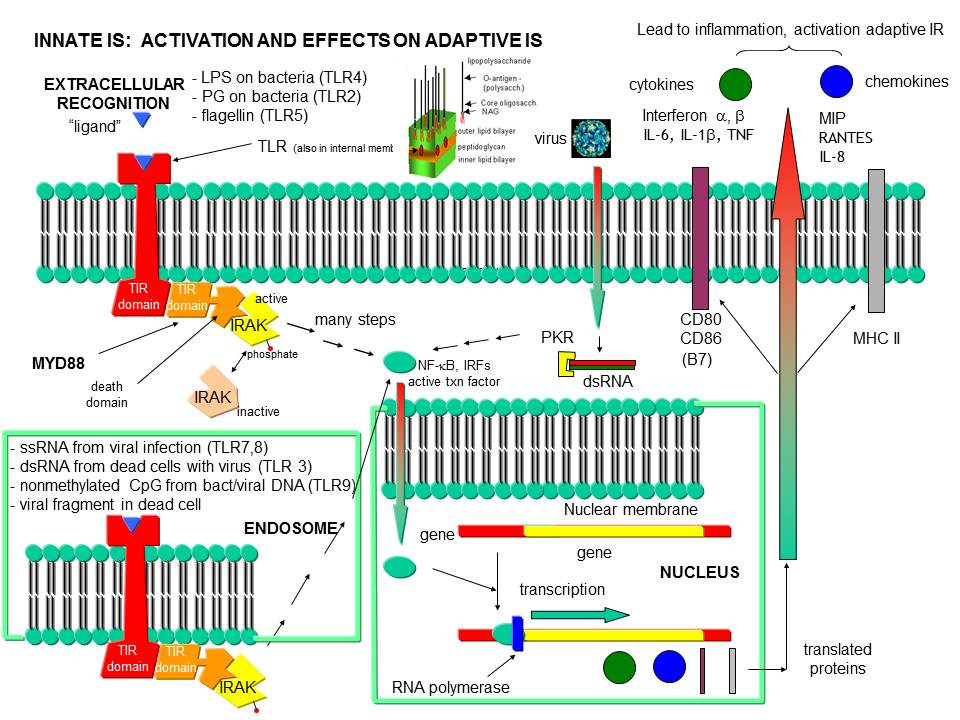
- Endosomal TLR3 Interaction with foreign RNA and DNA
- Recognition of PAMPs by TLRs
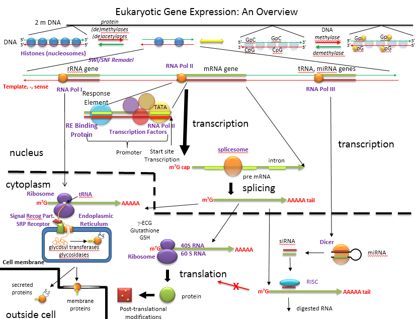
- An Overview of the Regulation of Gene Expression in Eukaroytes
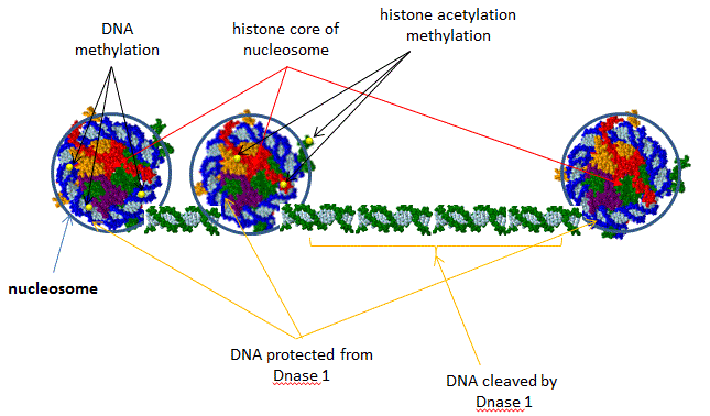
- DNase I Sensitivity of Nucleosome Bound and Free DNA
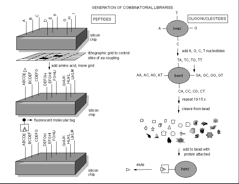
- Combinatorial Drug Development.
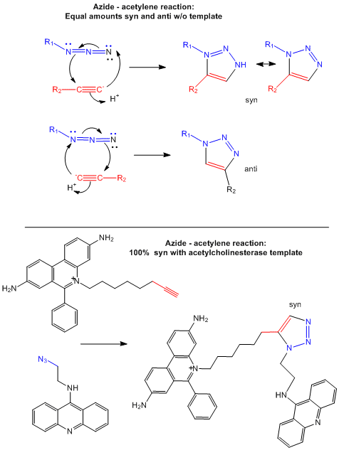
- Test tube and in situ synthesis of triazole inhibitors.
- Antisense RNA forms a dsRNA
- Cells can be made to make an antisense RNA within the cells
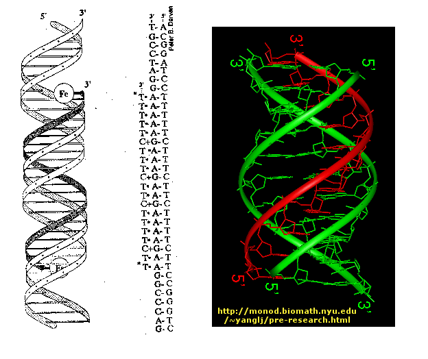
- Triple helix formation: binding to exposed H bond donors and acceptors of bases not involved in Watson-Crick H-bonding interactions.
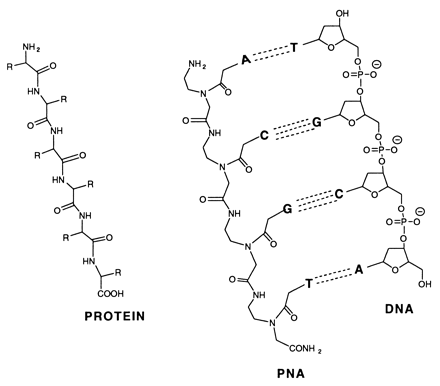
- Peptide:nucleic acid hybrids
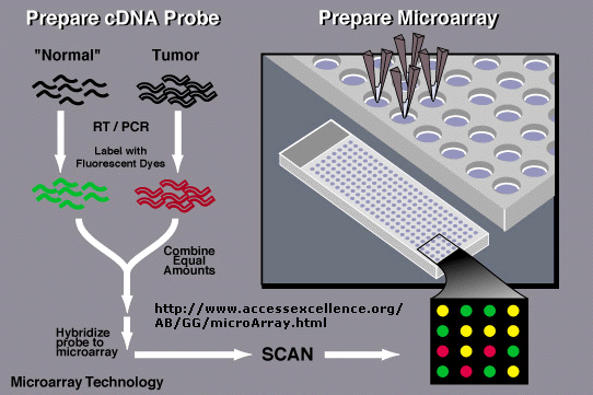
- Detection of Differential Gene Expression in Tumors and Normal Cells Using Microarray Chip
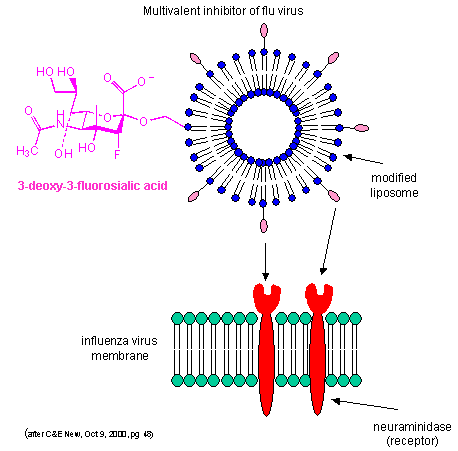
- Multivalent inhibitor of flu virus
- Multivalent Ligand Binding Models
Chapter 6: Transport and Kinetics
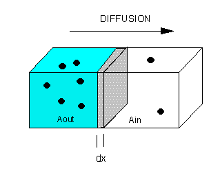
- Passive diffusion through a membrane - case 1: no receptor
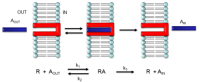
- Faciliated diffusion through a membrane - case 2: receptor-mediated
- Models for facilitated diffusion of glucose
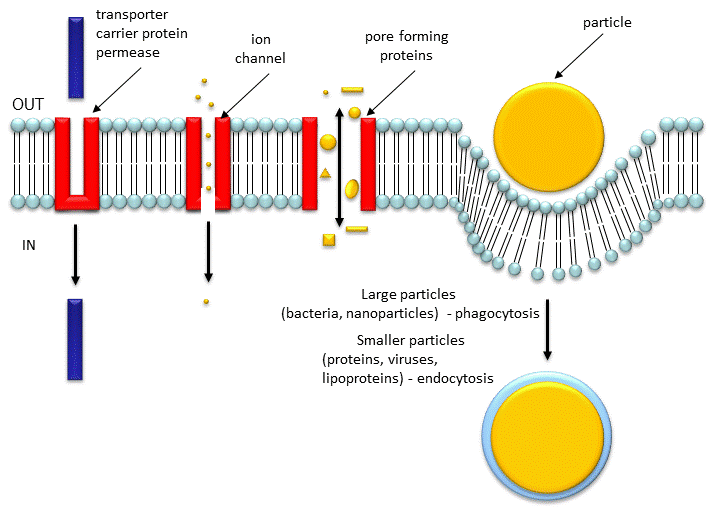
- Movement of Molecules and Particles Through Membranes
- Animation of ribitol diffusion through a glycerol channel using interactive molecular dynamics
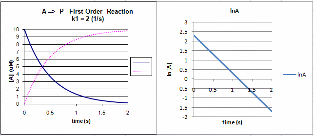
- First Order Reaction:
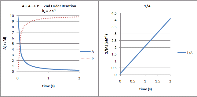
- Second Order Reaction:
- Reversible First Order Reactions:
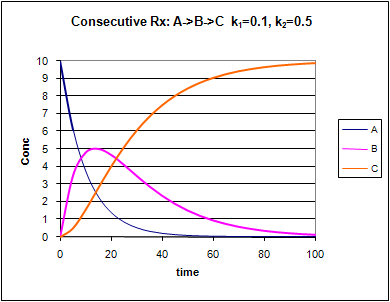
- Consecutive Irreversible First Order Reactions:
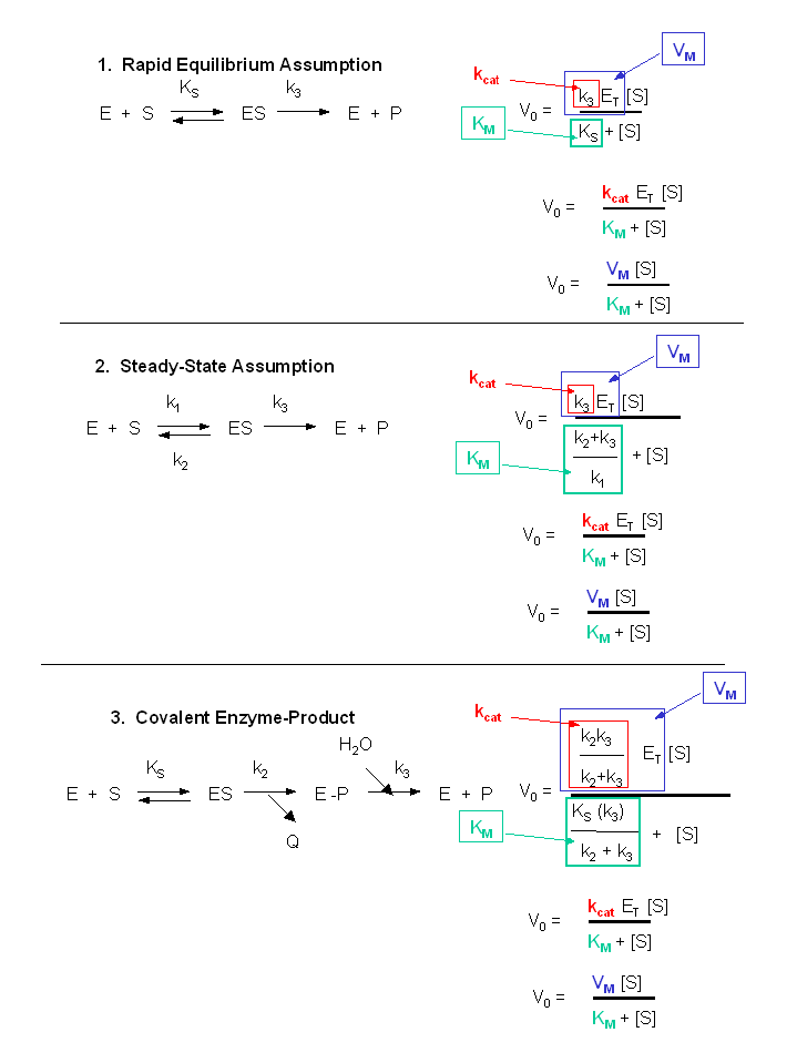
- Summary of vo vs S equations - Meanings of Km, kcat and Vm.
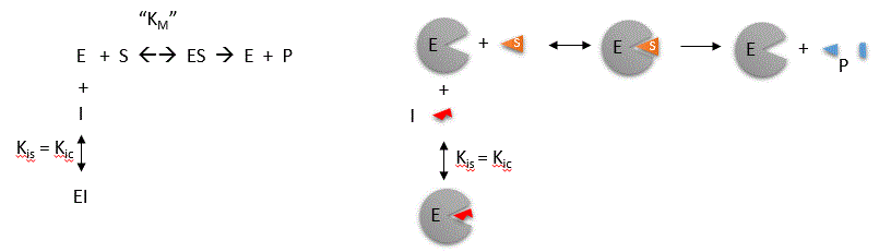
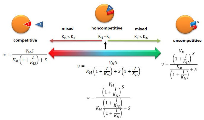
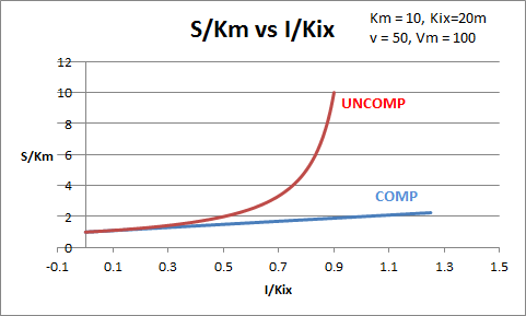
- Competitive inhibition
- Inhibition of Enzyme Activity - % Activity vs log [Inhibitor]
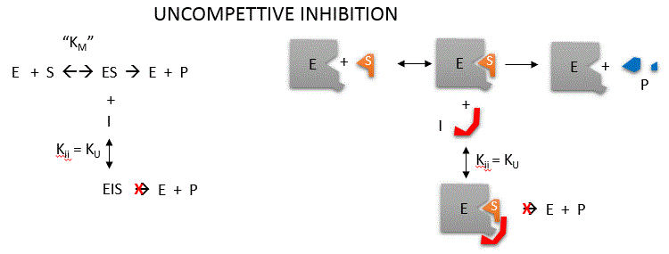
- Uncompetitive inhibition
- Noncompetitive inhibition
- Summary of Reversible Enzyme Inhibition
- Competitive and Uncompetitive Inhibition in vivo
- Agonist and Partial Agonists
- Antagonists: Competitive and Noncompetitive (Mixed)
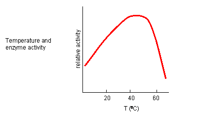
- Temperature and Enzyme Activity
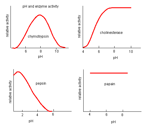
- pH and Enzyme Activity
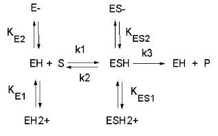
- Chemical equations showing the mechanism of pH effects on enzyme catalyzed reactions
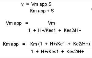
- Mathematic equations modeling pH effects on enzyme catalyzed reactions
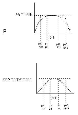
- Graphs of pH effects on kinetic constants for enzyme catalyzed reactions
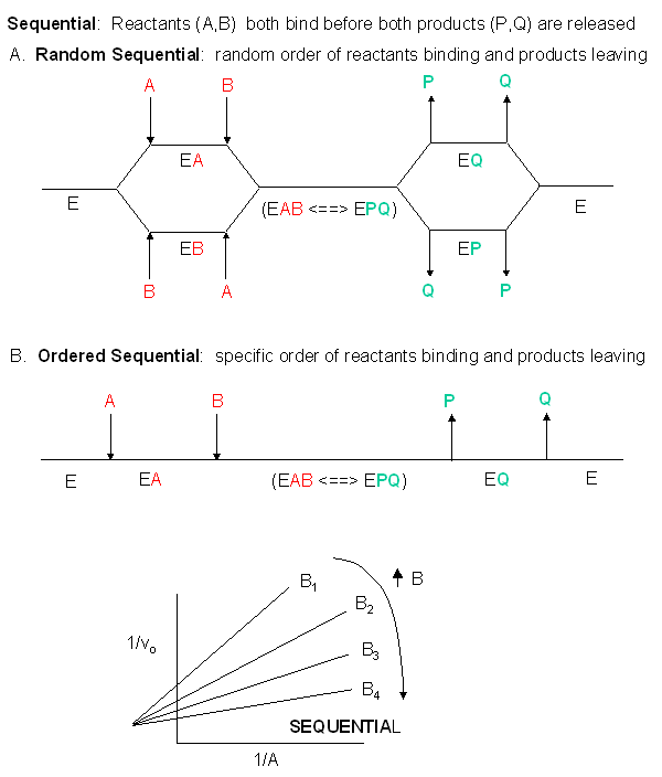
- Bisubstrate sequential kinetics
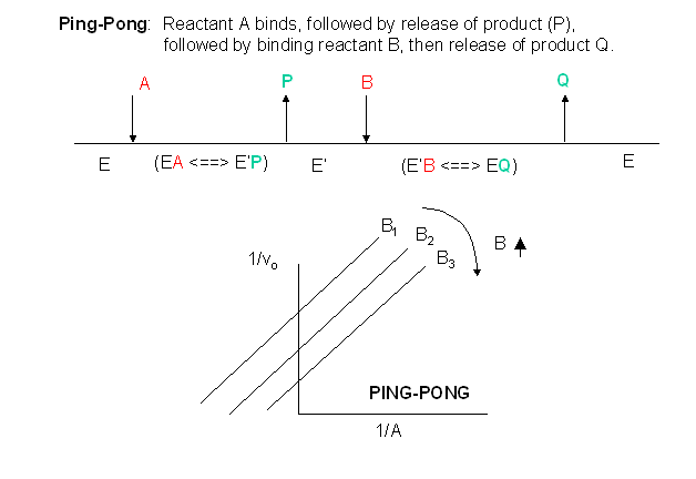
- Bisubstrate ping pong kinetics
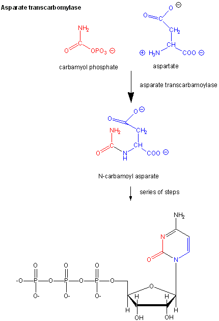
- Aspartate transcarbamoylase: reactions
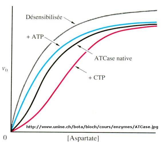
- Aspartate transcarbamoylase: Non Michaelis-Menten Kinetics
![Inhibition of Enzyme Activity - % Activity vs log [Inhibitor]](https://employees.csbsju.edu/hjakubowski/classes/ch331/transkinetics/inhibitionactivitylog.gif)
Chapter 7: Catalysis
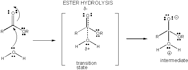
- Charge development in the transition state fore ester hydrolysis
- Mechanism of general acid catalysis
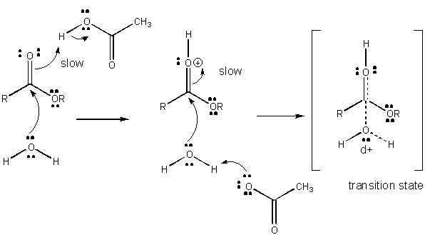
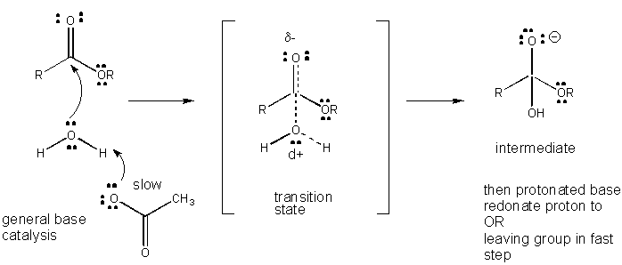
- Mechanism of general base catalysis
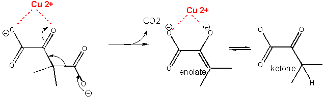
- Metal ion or electrostatic catalysis - stabilization of the transition state
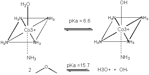
- Metal ion catalysis - decreasing the pKa of water
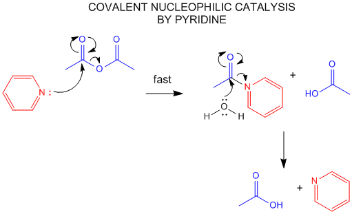
- Nucleophilic covalent catalysis by pyridine
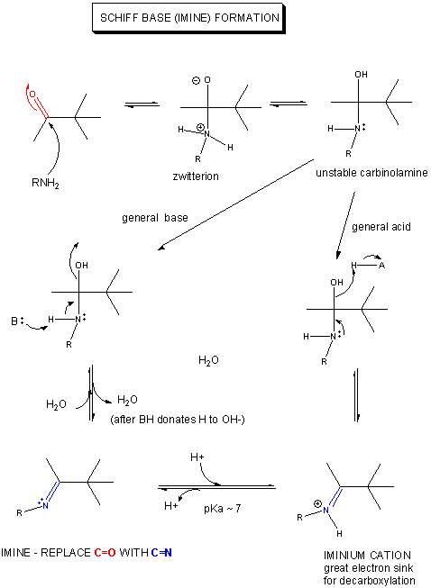
- Mechanism of Schiff base formation
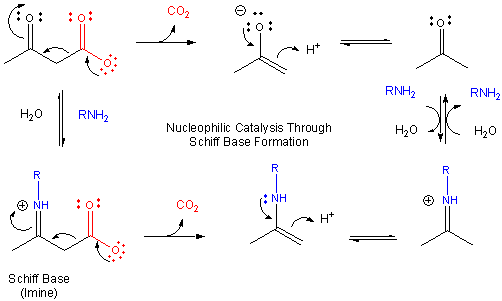
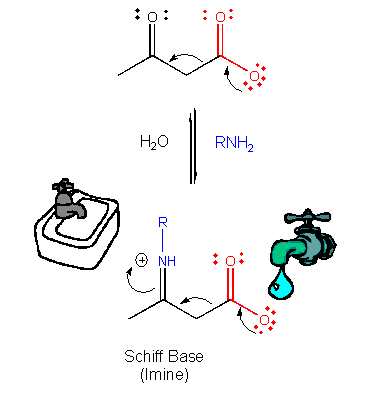
- Mechanism of nucleophilic catalysis by amines - Schiff base formation .
- Electron Flow: Source to Sink
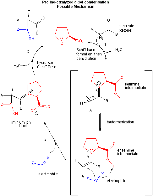
- L-Pro catalysis of an aldol condensation: possible mechanism
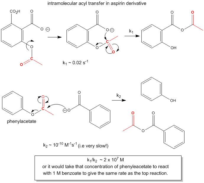
- Acyl transfer in aspirin derivatives
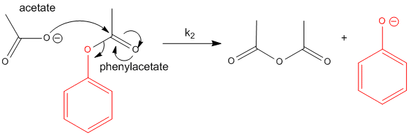
- Reaction of acetate with phenylacetate
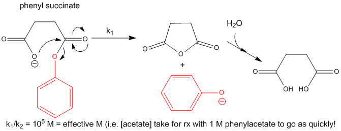
- Intramoleuclar reactions of phenylsuccinate
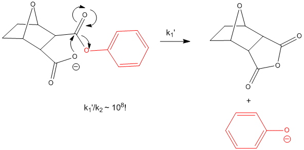
- Intramolecular reactions of bicyclic phenylcarboxlate
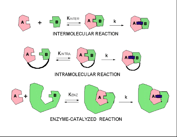
- Three kinds of reactions, intermolecular, intramolecular, and enzyme-catalysed
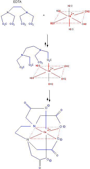
- Binding of Ca2+ and EDTA
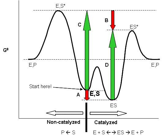
- Enyzmes bind the transition state more tightly than the substrate
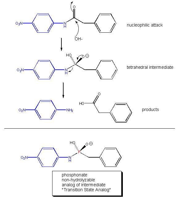
- Phosphonamides: transition state analogs
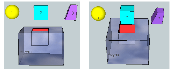
- 3D model for binding substrate, transition state, and product to an enzyme
- Carboxypeptidase: mechanism of peptide bond cleavage
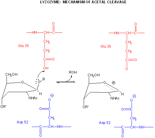
- Lysozyme: mechanism of acetal cleavage
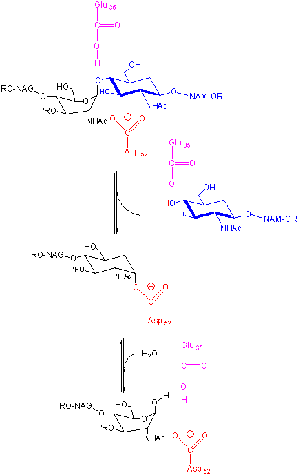
- Lysozyme: alternative mechanism
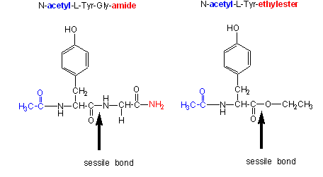
- Amide/ester substrates for chymotrypsin
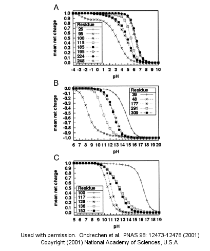
- Theoretical microscopic titration curves
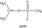
- Diisopropylphosphofluoridate
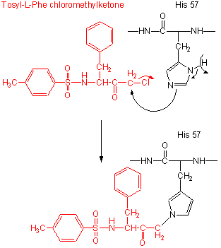
- Tos-L-Phe-chloromethyl ketone
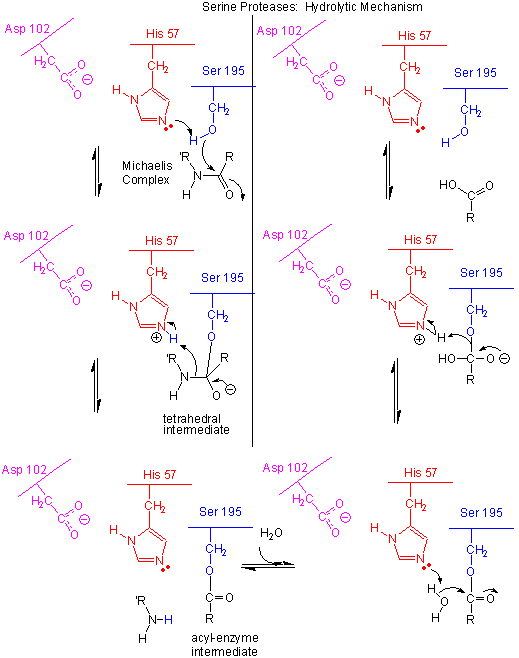
- Serine Protease Mechanism
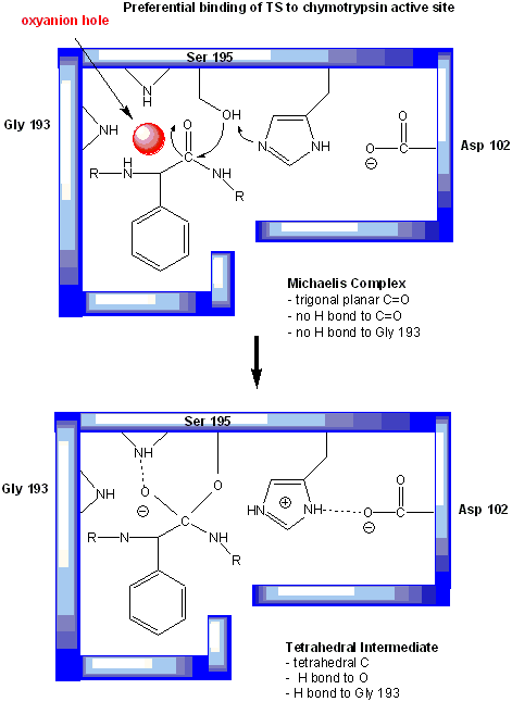
- Oxyanion hole in serine proteases: TS stabilzation
- Sarin
- Cleavage of beta amyloid precursor protein: protease and cofactors
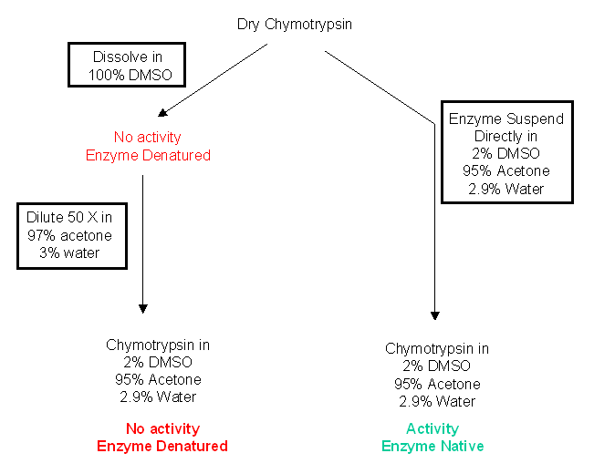
- Chymotrypin activity - DMSO to acetone
- Ribozyme tertiary structures
- Ribozyme: mechanisms of catalysis
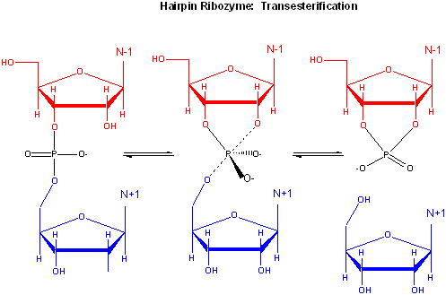
- Self-cleavage in the hairpin ribozyme
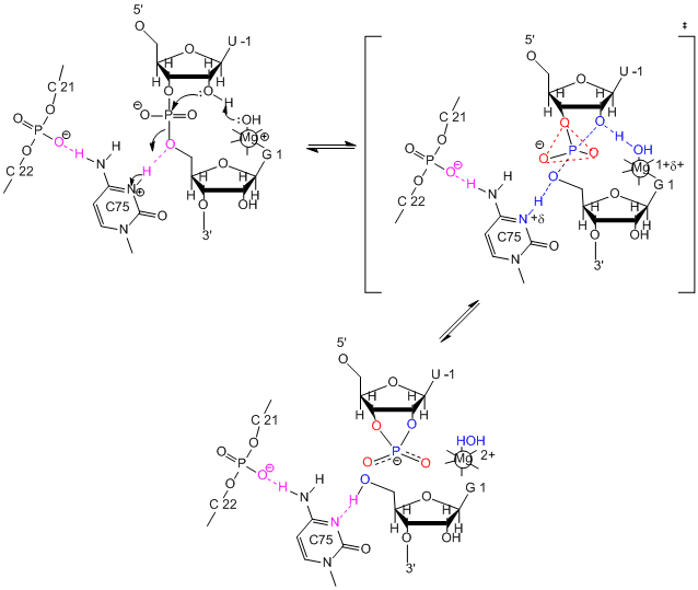
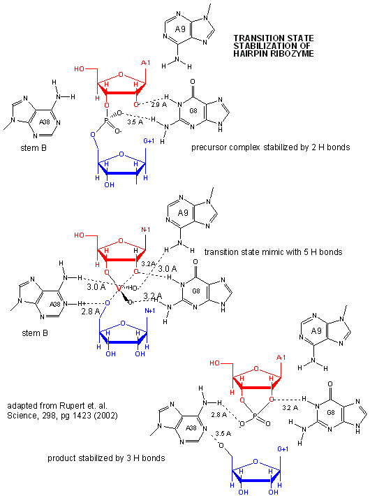
- Active Site of Hairpin Ribozyme: Transition State Binding
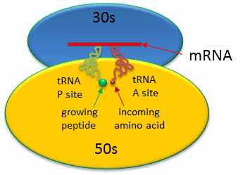
- Prokaryotic Ribosme - P and A sites
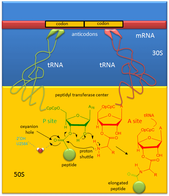
- Mechanism Peptide Bond Formation by the Ribosome
Chapter 8: Oxidation/Phosphorylation
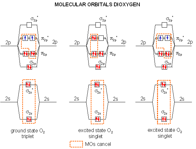
- Dioxygen: MO's for ground state triplet and excited state singlet dioxygen
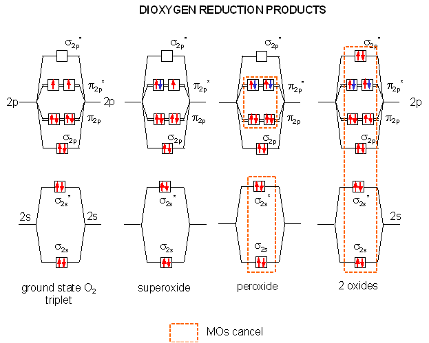
- Dioxygen reduction: MO's and products
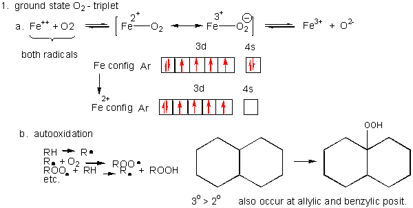
- Triplet O2 - Ground State chemistry
- Unsaturated fatty acids are extra reactive at the methylene C that separate the double bonds
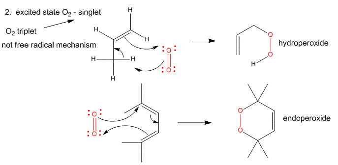
- Single O2 - Excited State chemistry
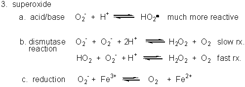
- Superoxide
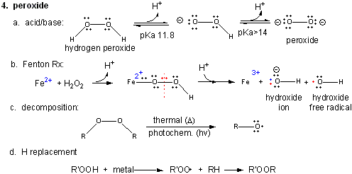
- Peroxide chemistry
- Hydroxyl free radical
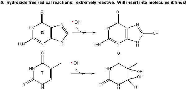
- Oxidative Modification of DNA
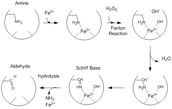
- Oxidative Modification of Proteins
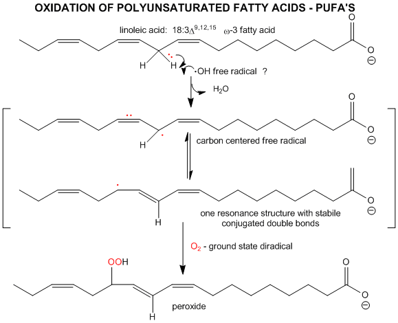
- Oxidative Modification of Lipids
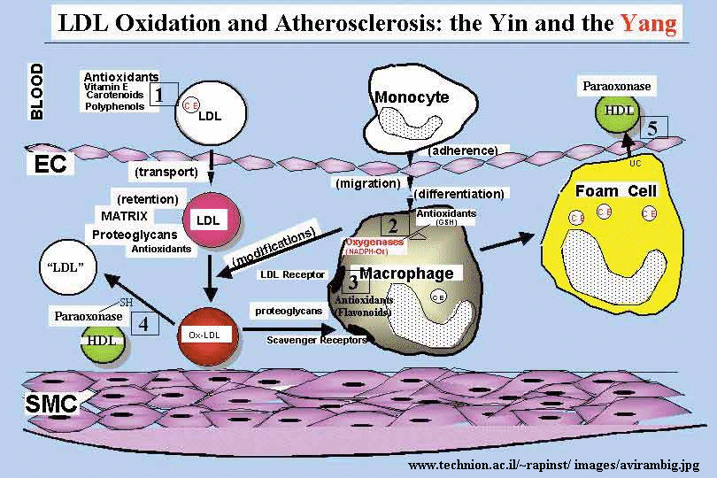
- Oxidized LDL uptake
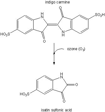
- Conversion of indigo carmen to isatin sulfonic acid
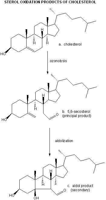
- Cholesterol/ozone reaction
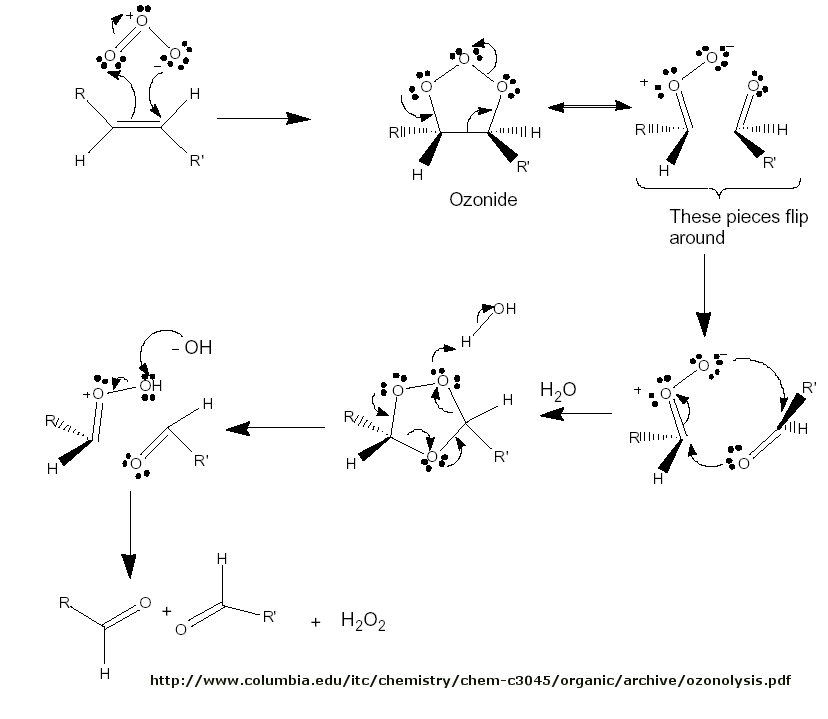
- Ozonolysis mechanism
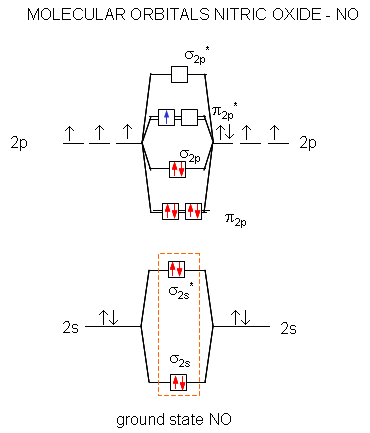
- Molecular Orbitals: NO
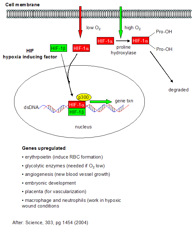
- Cell response to hypoxia
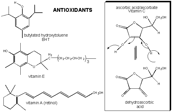
- Antioxidant vitamins (E, C, A)
- Mechanisms of combustion reactions
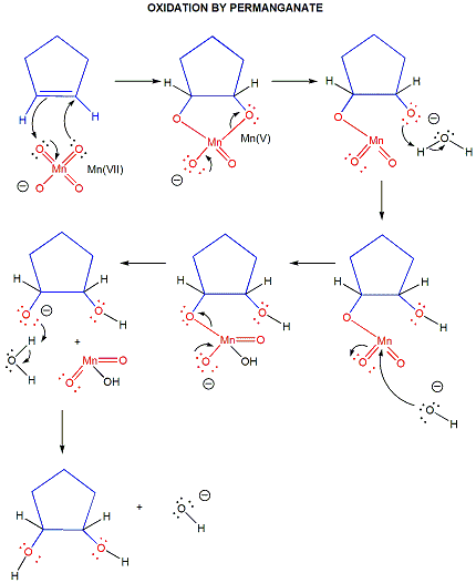
- Permanganate
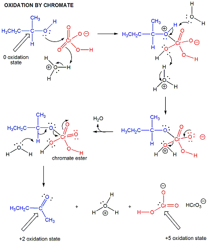
- Chromate
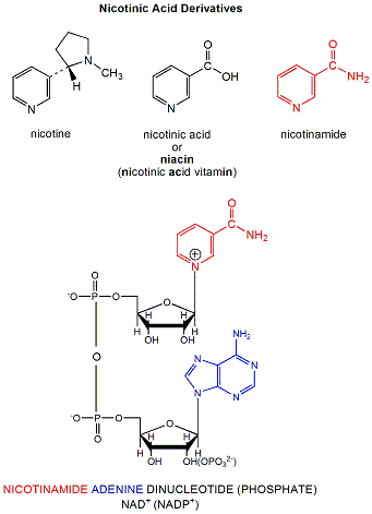
- NAD+ is a derivative of nicotinic acid or nicotinamide
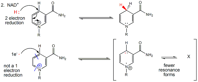
- All NAD+/NADH reactions in the body involve 2 electron hydride transfers
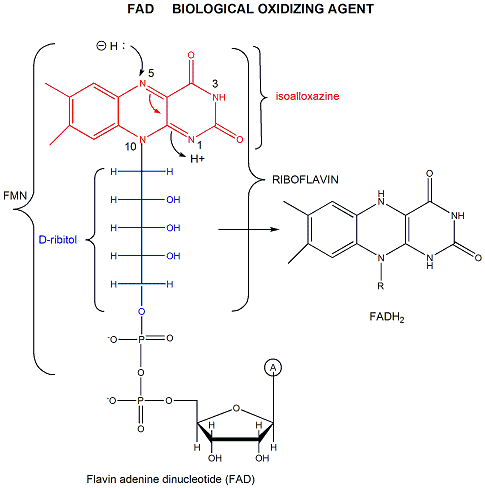
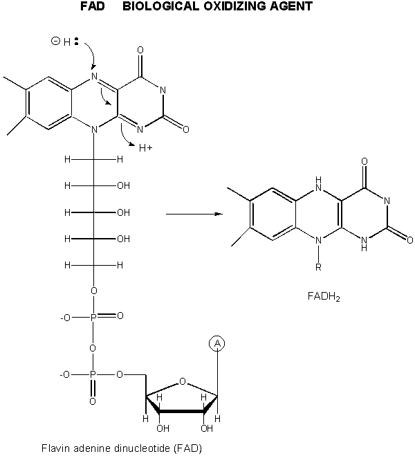
- derivatives of riboflavin.
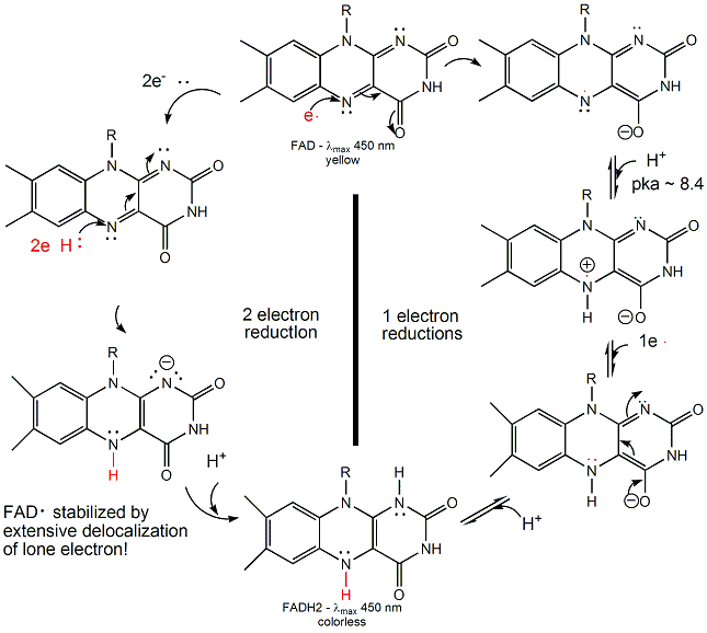
- FAD/FADH2 can undergo 1 OR 2 electrons transfers
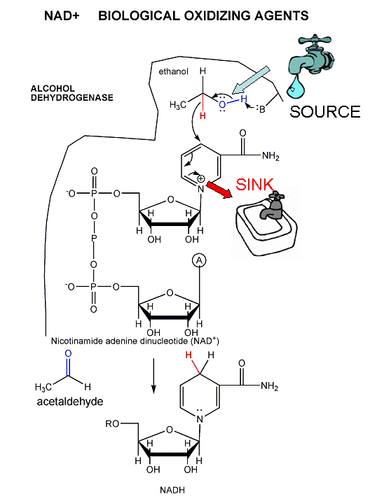
- Oxidation of ethanol to acetaldehyde by alcohol dehydrogenase.
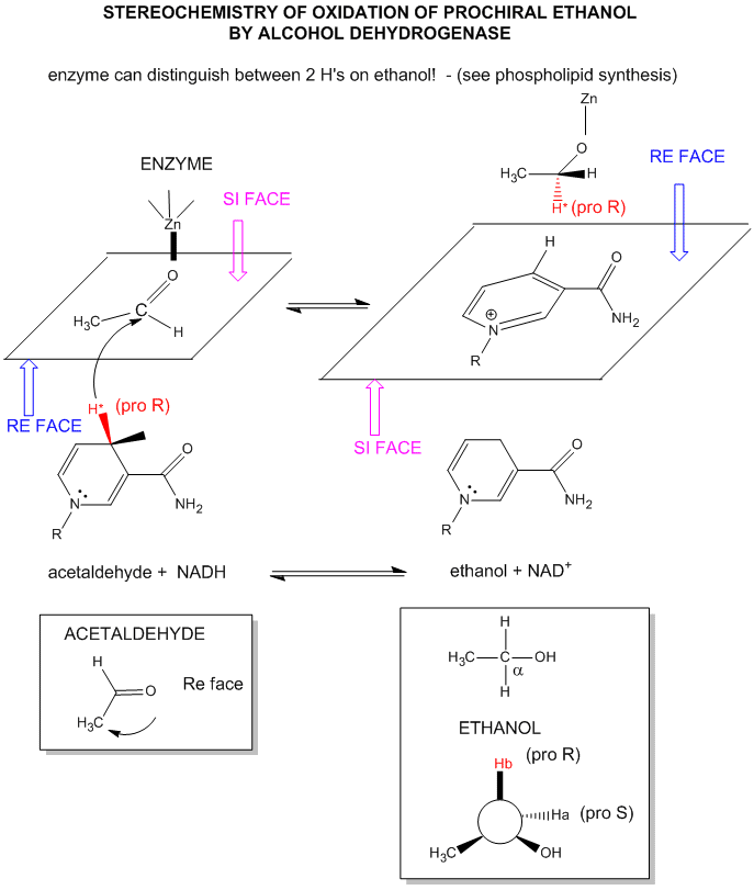
- Stereochemistry of NAD+/NADH redox reactions with alcohol dehydrogenase
- FAD oxidation reactions: mechanisms
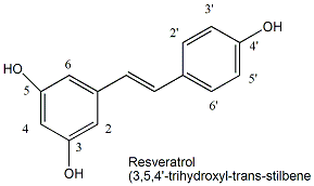
- Structure of resveratrol: activator of Sir2 deacetylase activity
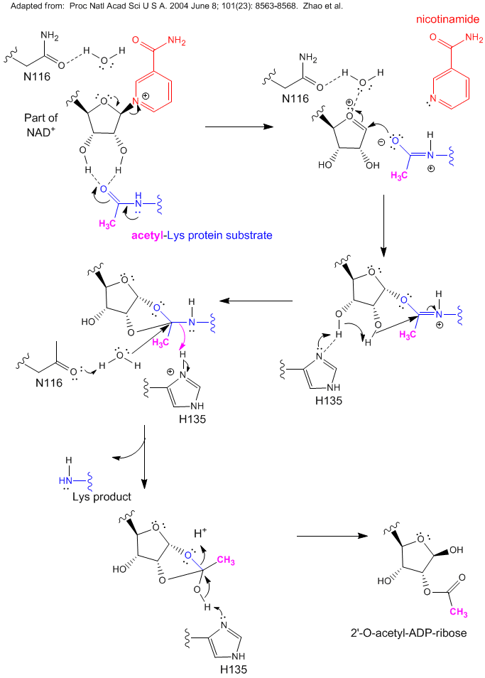
- Reaction of NAD+ and Acetyl-Lys side chains: possible catalytic mechanism for yeast Sir2
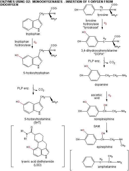
- Trp and Tyr chemistry
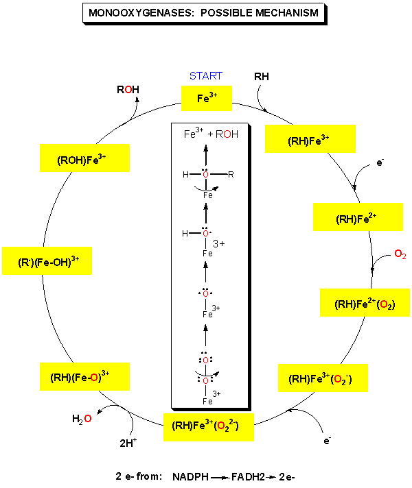
- Monooxygenases: possible mechanism
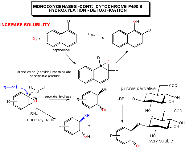
- Cytochrome p450's: possible mechanism
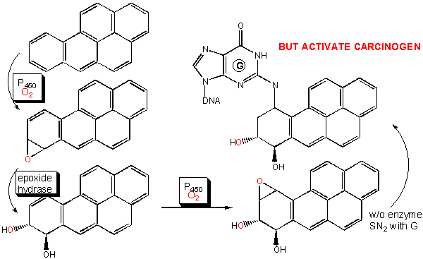
- Cytochrome p450's: activation of carcinogens
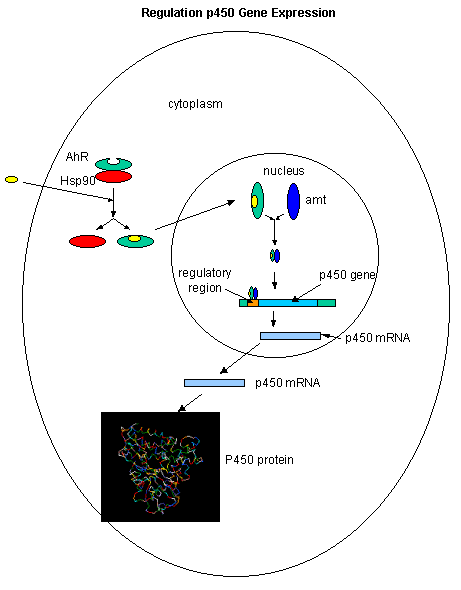
- Inducible on exposure to nonpolar aromatic molecules such as dioxin.
- Reaction Pathyway of P450cam
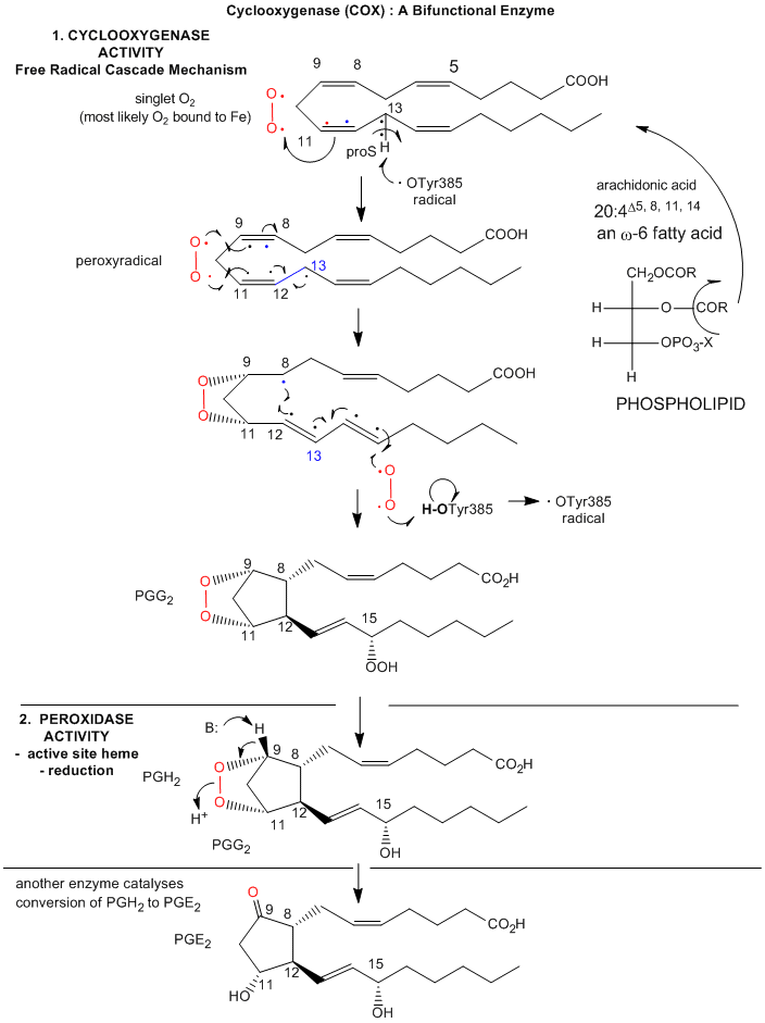
- Arachadonic Acid Bound to the Active Site of Mouse Cyclooxygenase
- Prostatglandin Synthesis: Possible Mechanism
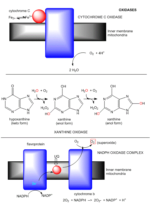
- Oxidases: Examples
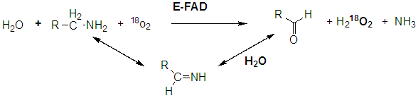
- Monoamine oxidases
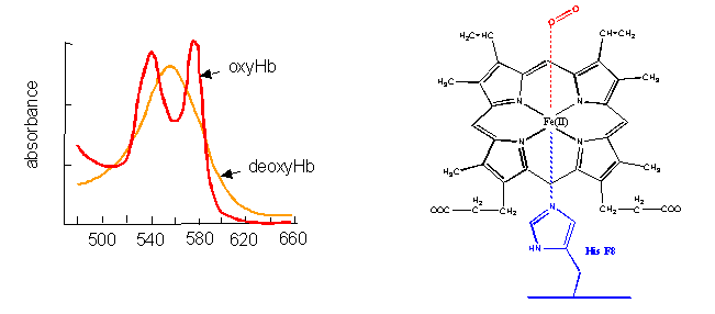
- Hemoglobin (and myglobin) serve as carriers of dioxygen.
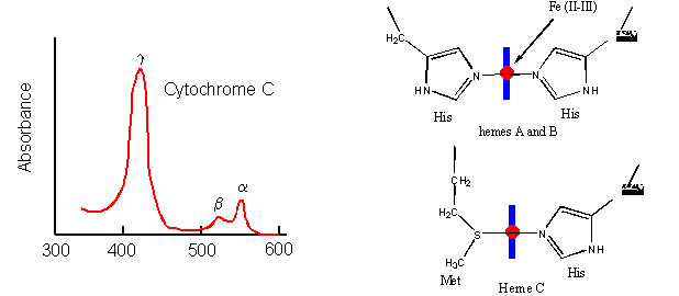
- Cytochrome C
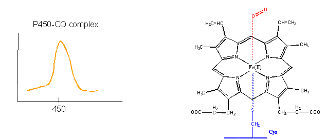
- cytochrome P450's
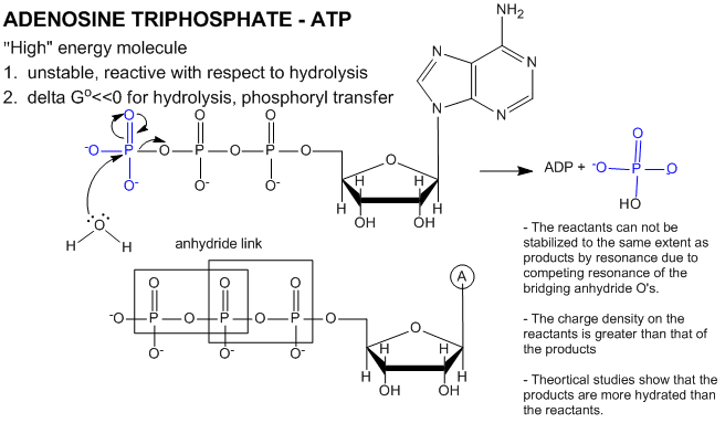
- Structure and hydrolysis of ATP
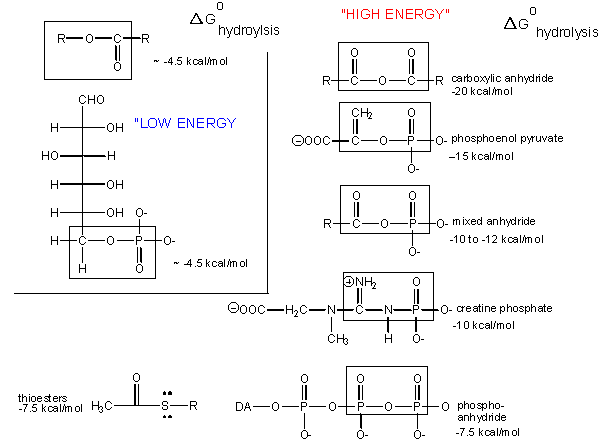
- High energy molecules
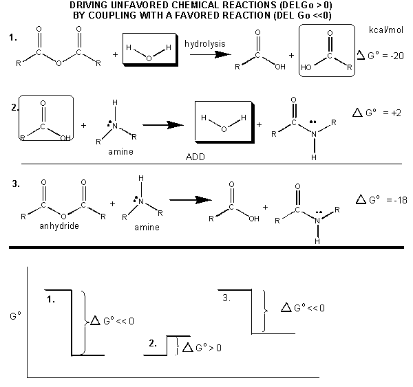
- Mechanism: coupled synthesis of a carboxylic amide
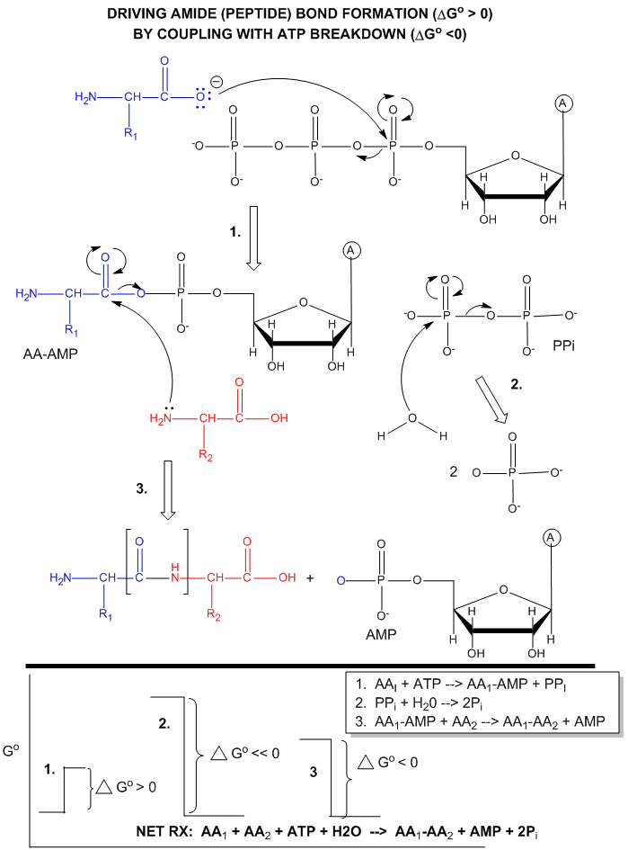
- Mechanism: ATP-dependent peptide bond synthesis
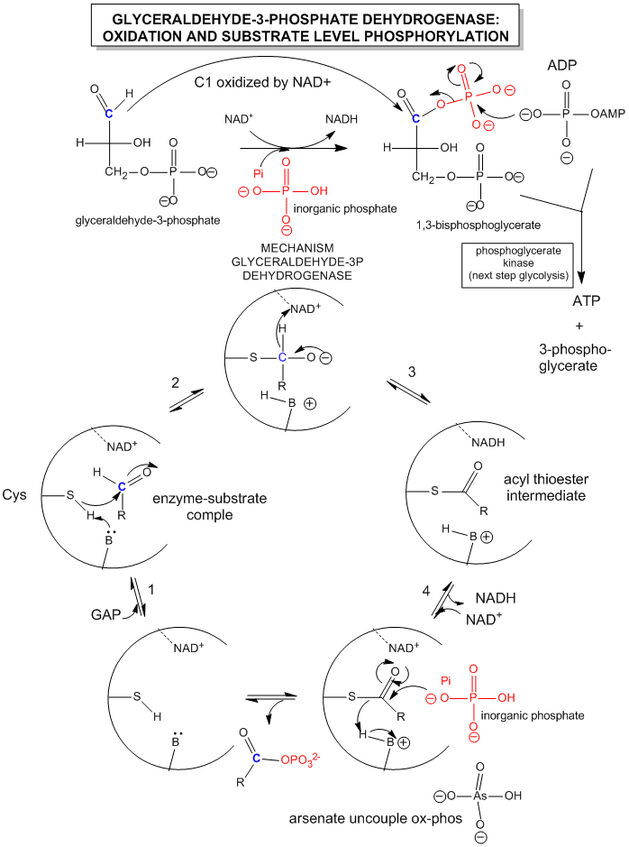
- Mechanism of OX-PHOS in glycolysis: glyceraldehye-3-phosphate dehydrogenase and 3-phosphglycerate kinase
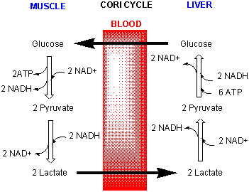
- Cori Cycle
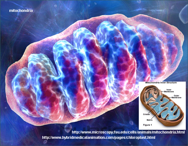
- Mitochondria
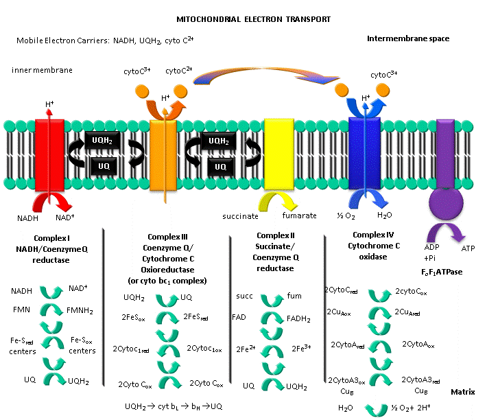
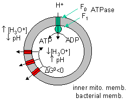
- ELlectron transport and proton gradient formation in the mitochondria
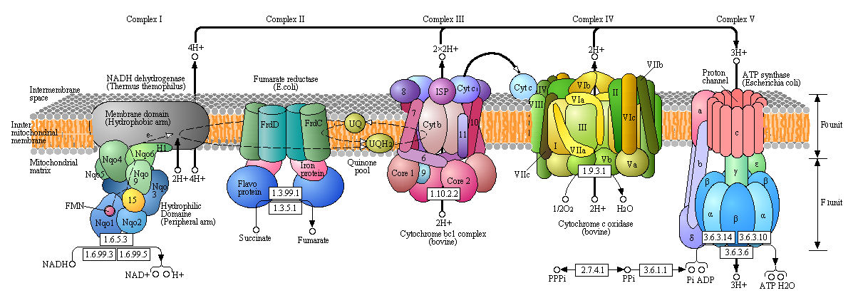
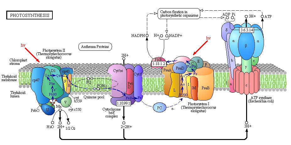
- Detailed View of Oxidative Phosphorylation from Kanehisa Laboratories and the KEGG project: www.kegg.org
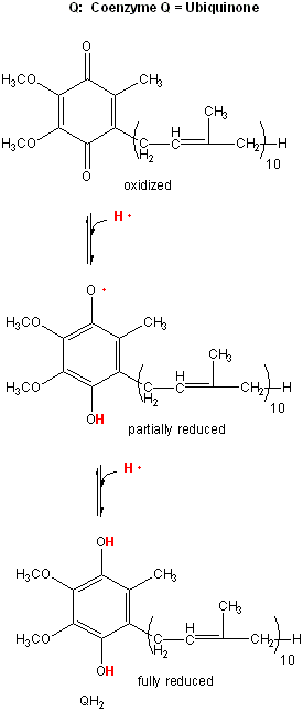
- Lipophilic electron carrier, ubiquinone, UQ
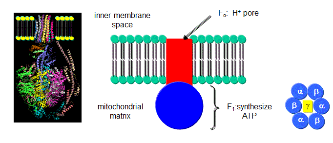
- FoF1ATPase, also called ATP synthase,
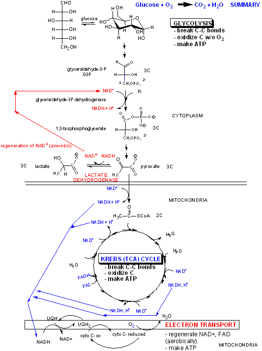
- Overview of metabolism: Aerobic and Anerobic Generation of NADH, Regeneration of NAD, and Coupling Oxidation/Phosphorylation
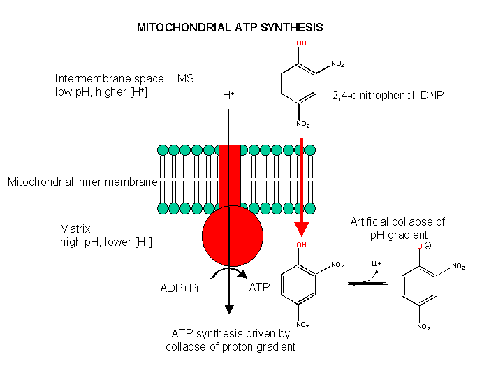
- Uncoupling Aerobic Ox/Phos
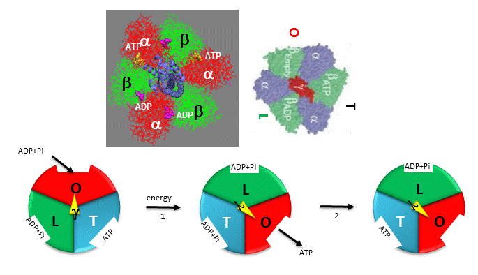
- Boyers three-state conformational model (L-O-T) for ATP synthesis.
- fluorescein-labeled protein filament called actin to the g subunit
- Smaller colloidal gold bead (40 nm diameter) with less frictional resistance
- Rotation rate becomes continuous and saturates
- Coupling Proton Flow in F0 to Conformation Change
- ATP synthase Complex
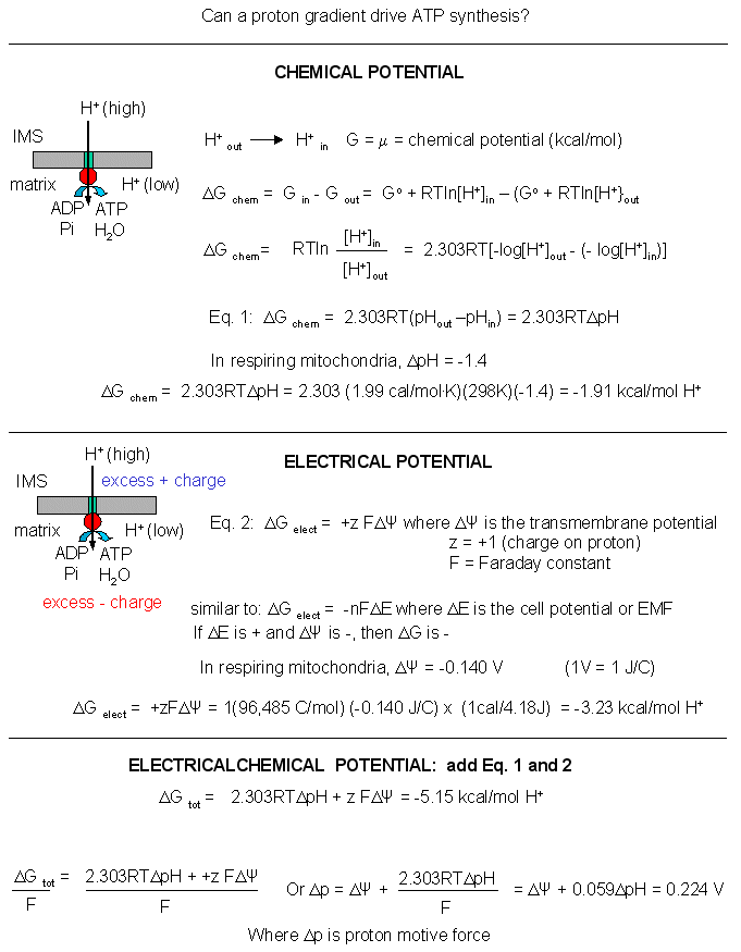
- A simple mathematical derivation: Electrochemical Potential
- Two different proton translocating methods
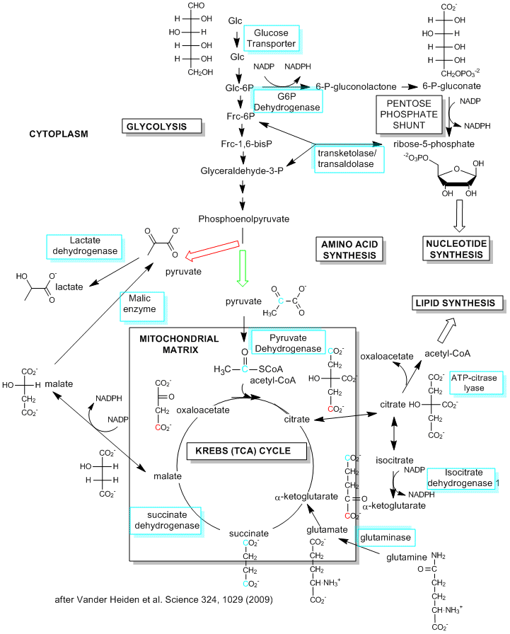
- Metabolic Pathways in Proliferating Cells
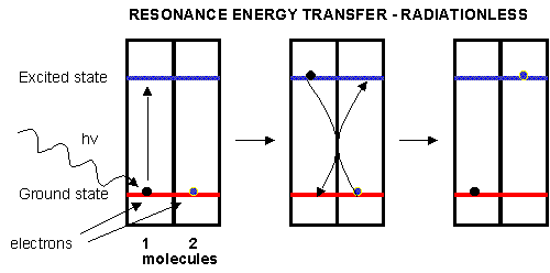
- Resonance energy transfer
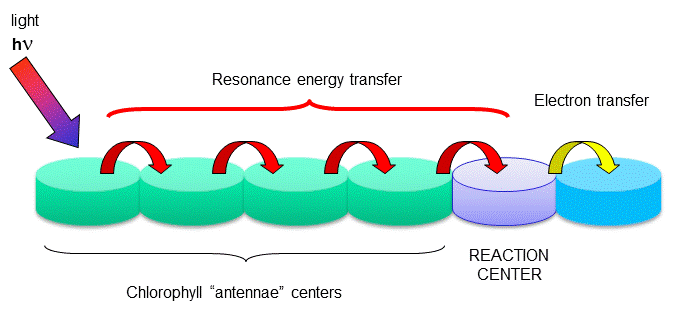
- Reaction centers
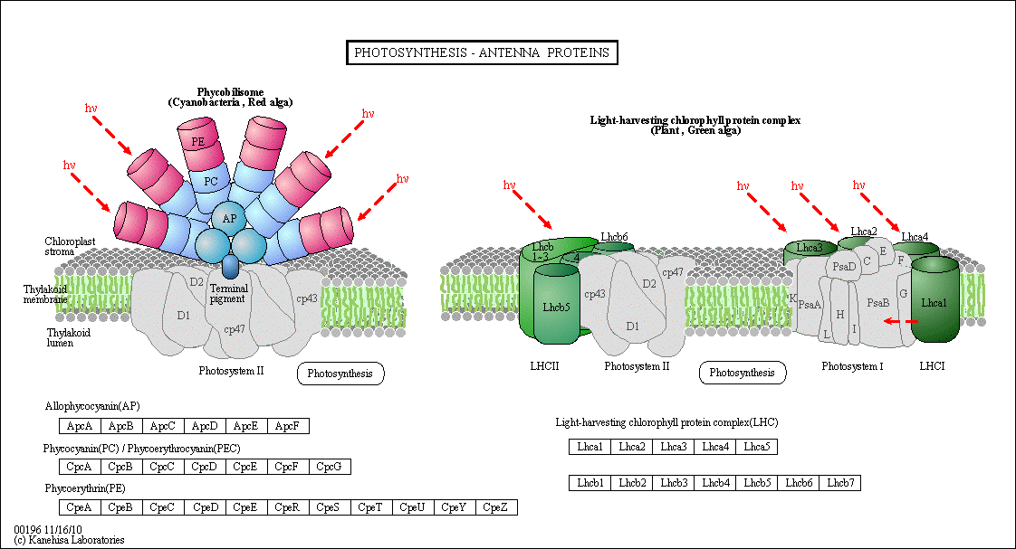
- Antennae Proteins
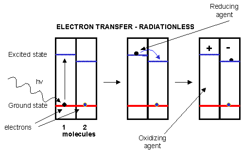
- Electron transfer
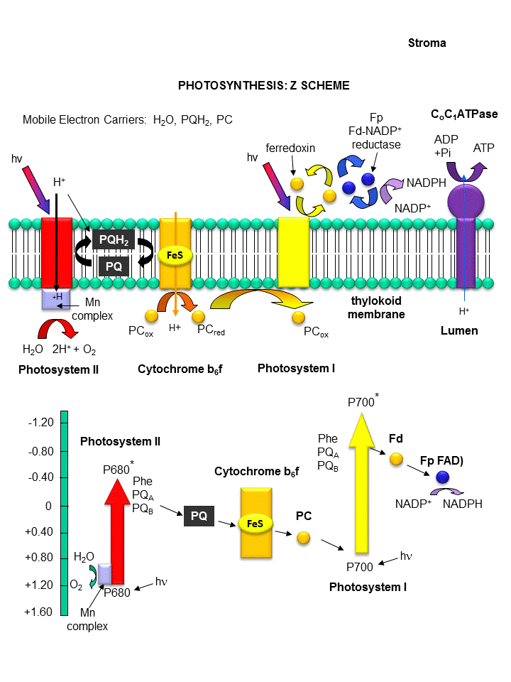
- Light reaction of photosynthesis
- Detailed View of Light Reaction of Photosynthesis
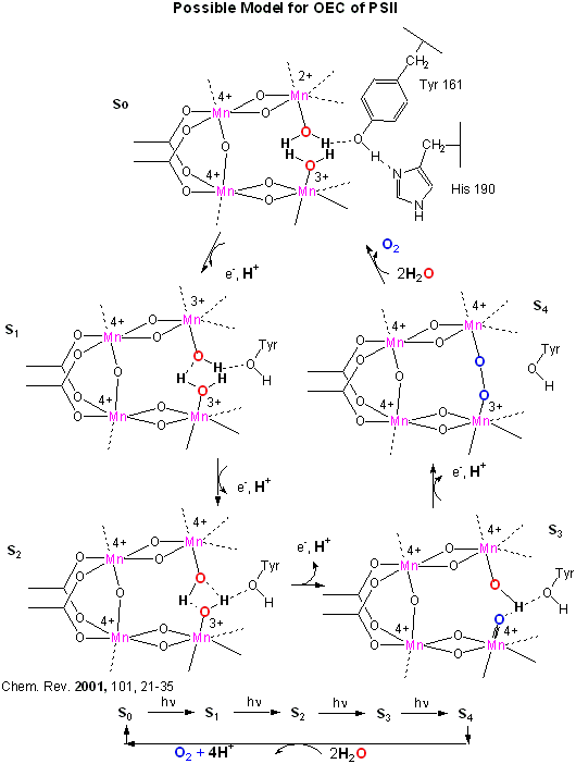
- OEC - Mechanism of Water Oxidation
Chapter 9: Signal Transduction
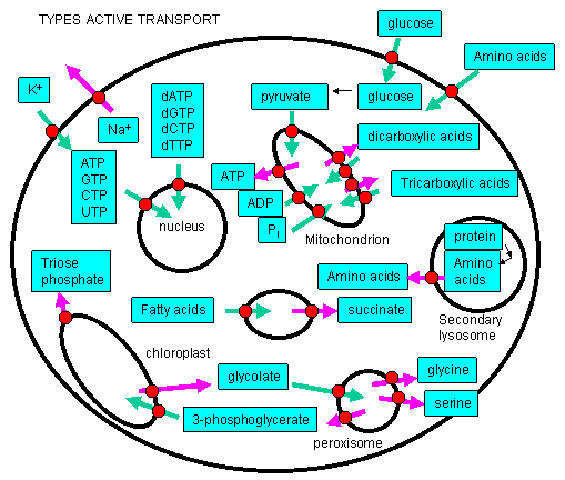
- Many species are transported into the cell or into intracellular organelles against a concentration gradient
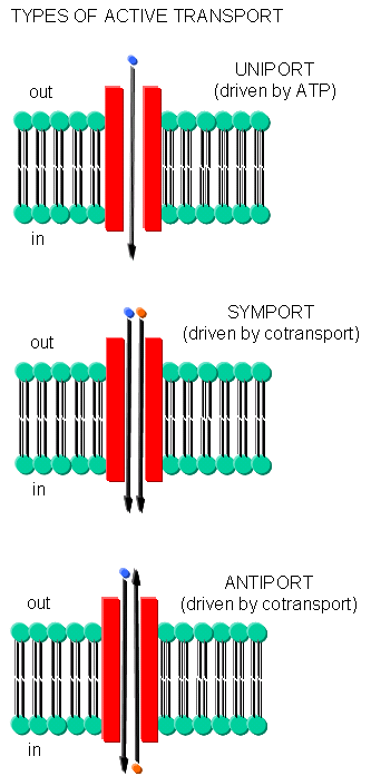
- Types of Active Transport
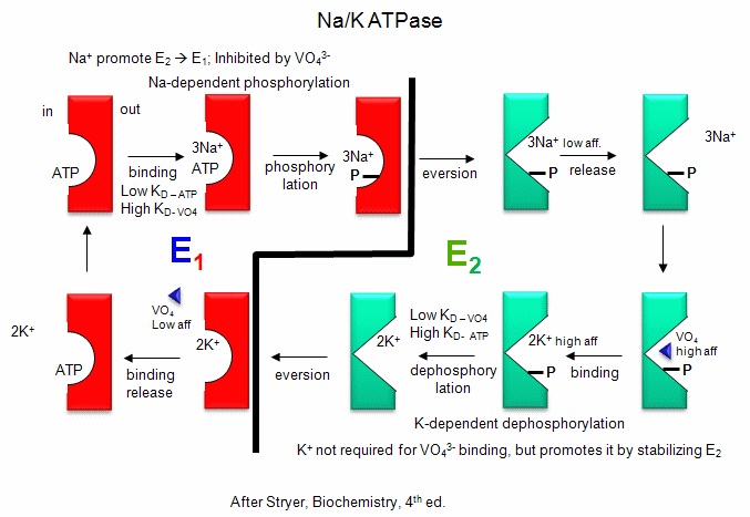
- Na-K-ATPase Model
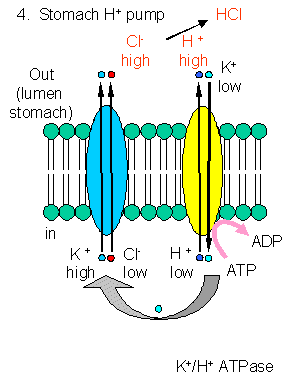
- HCl production in stomach
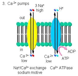
- Transport of calcium ions
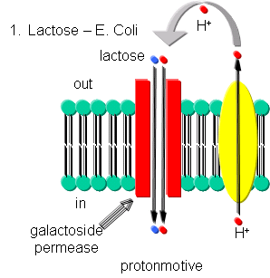
- Lactose Transport
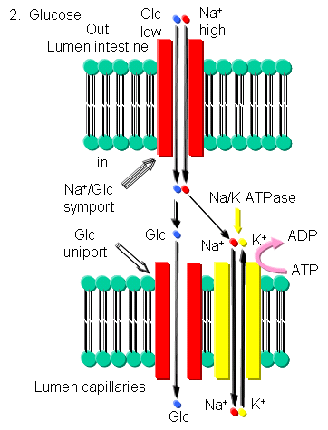
- Glucose Transport
- Proton transfer in bacteriorhodopsin
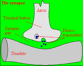
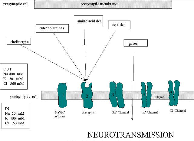
- neurotransmitter into the synapse between the cells
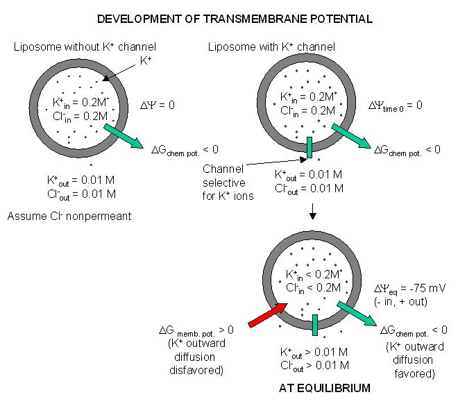
- Visualizing the transmembrane potential in K+ loaded vesicles + a nongated K+ channel.
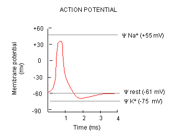
- Depolarization of transmembrane potential
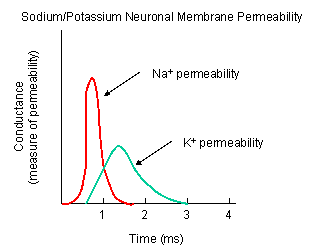
- Na and K permeabilities during depolarization
- Five membrane proteins
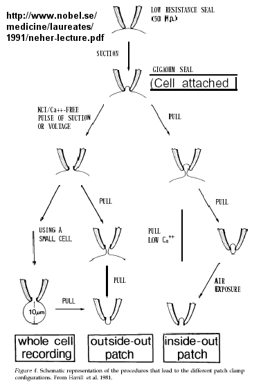
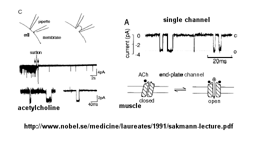
- Patch clamp technique
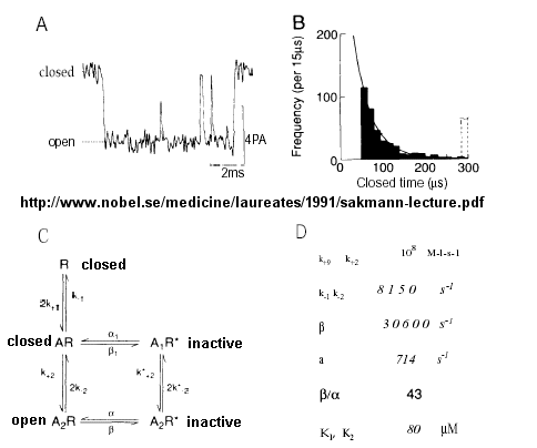
- Opening and closing of single channels
- Mechanism for gating of the acetylcholine channel
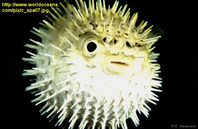
- Puffer fish
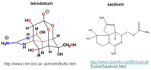
- Tetrodoxin and Saxitoxin - Structures
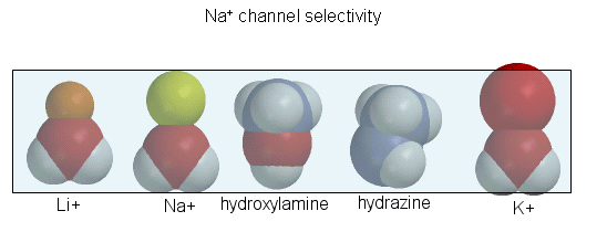
- Ion selectively of the sodium channel
- Electrostatics of the K+ channel
- K+ Channel from Steptomyces lividans
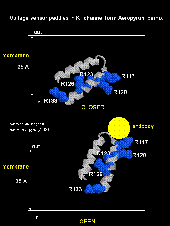
- S4 paddle voltage sensor of the voltage-dependent K+ channel from Aeropyrum pernix.
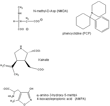
- Glutamate receptor ligands
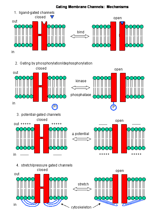
- Mechanisms of Channel Gating
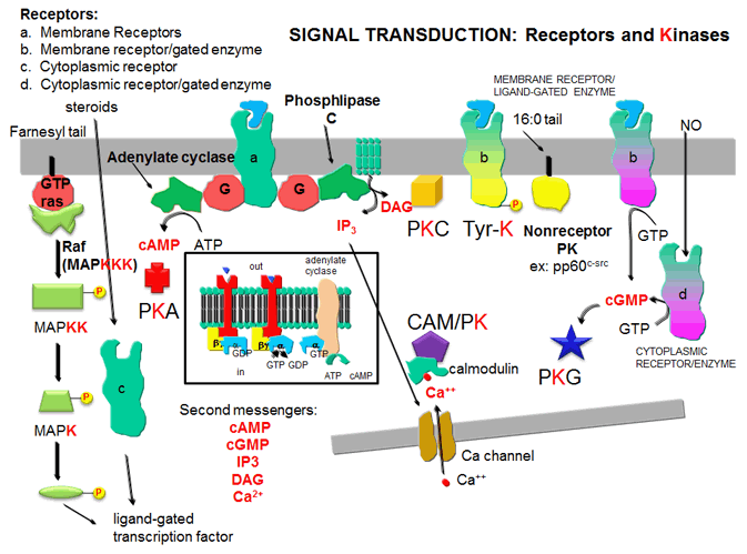
- five major protein kinase
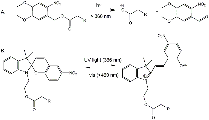
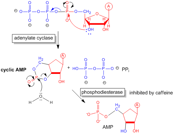
- cyclic AMP
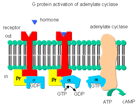
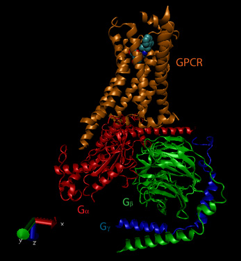
- G proteins and adenylate cyclase activation
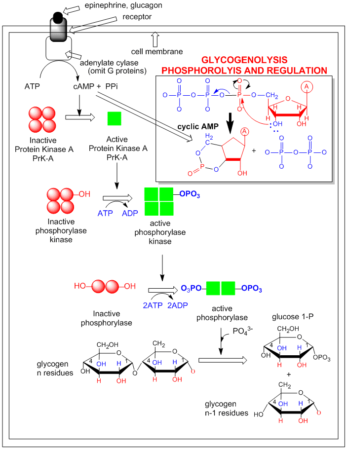
- Activation of glycogen phosphorylase through activation of PKA.
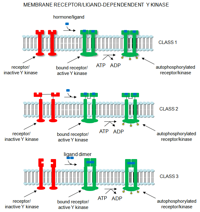
- Receptor/Ligand-Dependent Protein Kinases
- Beta-andrenergic:Gs complex
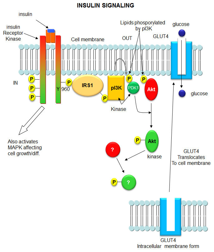
- Initial Signaling on Binding of Insulin to the Insulin Receptor
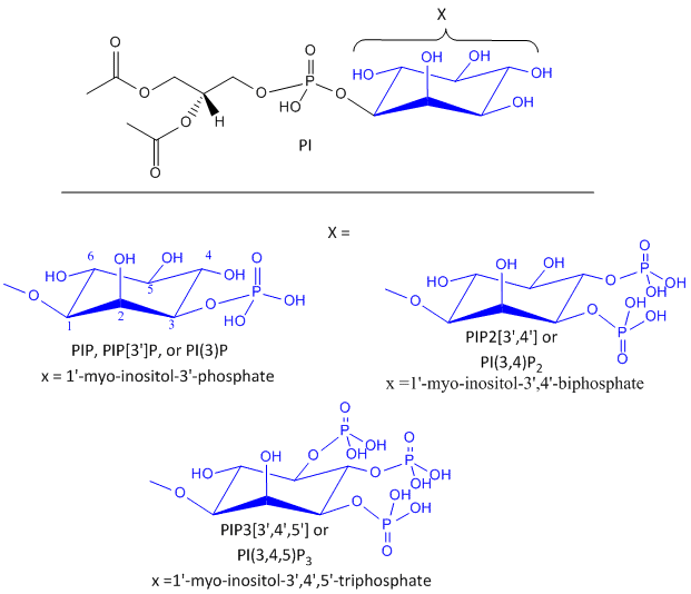
- Phosphorylated Phosphatidylinositol derivatives
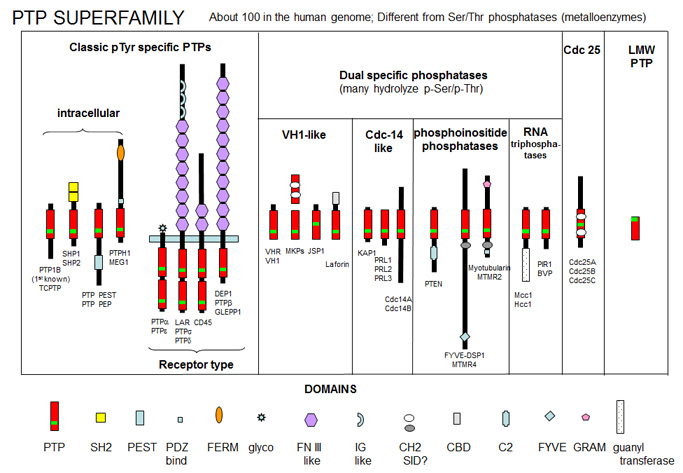
- PTP Super Family
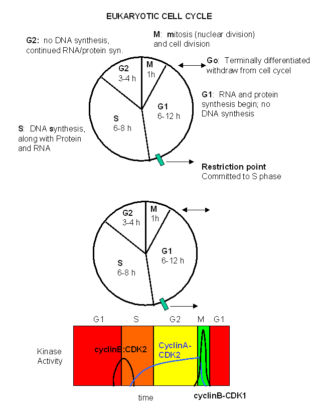
- Cell Cycle
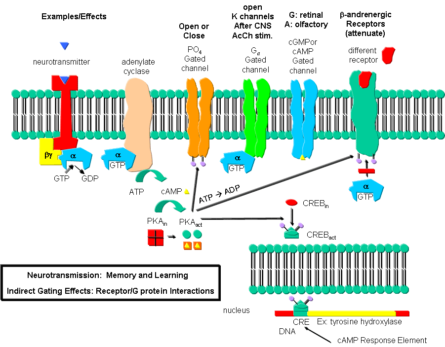
- Neurotransmission: Gating through G Linked Receptors
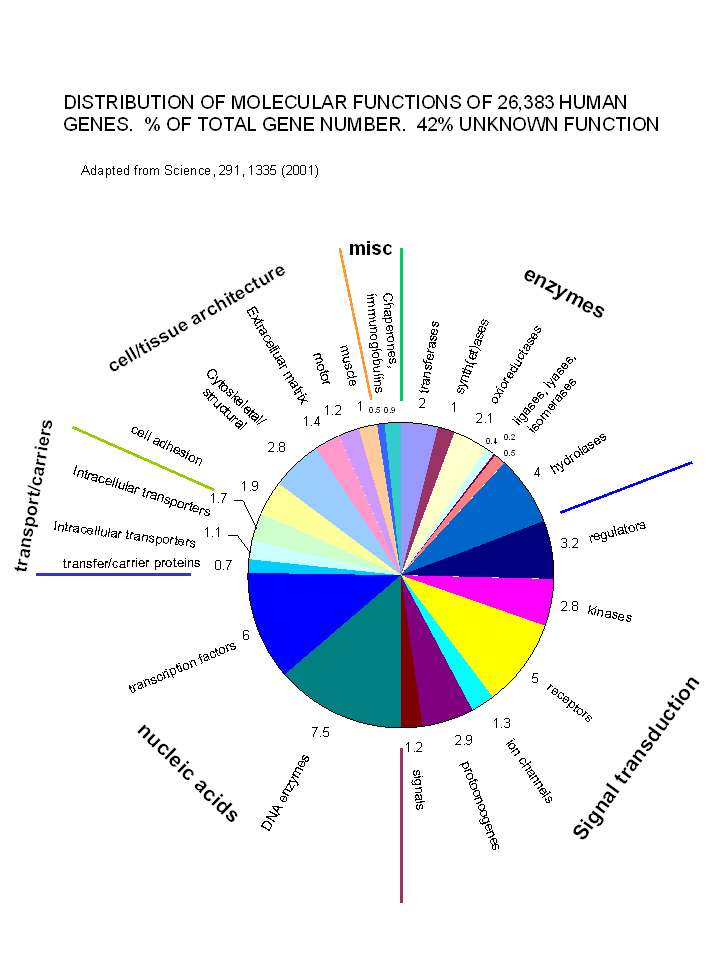
- Distribution of Molecular Functions of 26,383 Genes
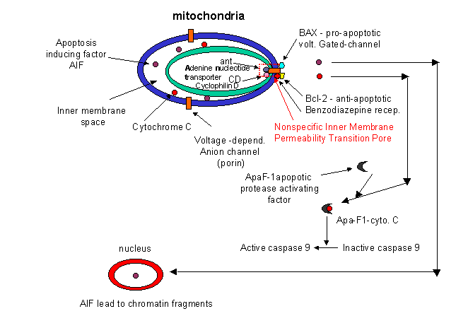
- Mitochondria Changes During Apoptosis
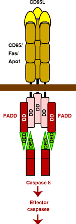
- CD95:CD95L Interactions
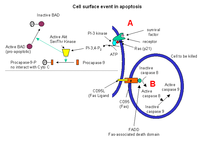
- Cell Receptor-Mediated Apoptosis
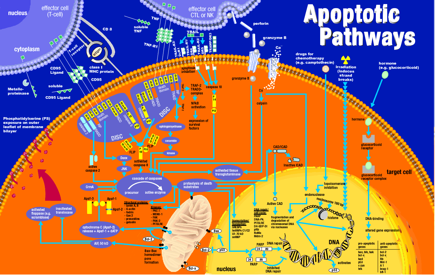
- Apoptosis Wall Chart
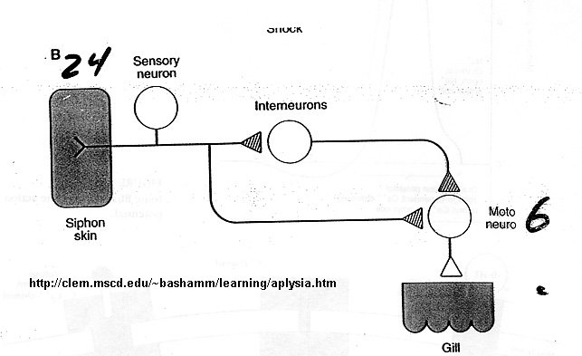
- Aplysia sensory cells in Habituation
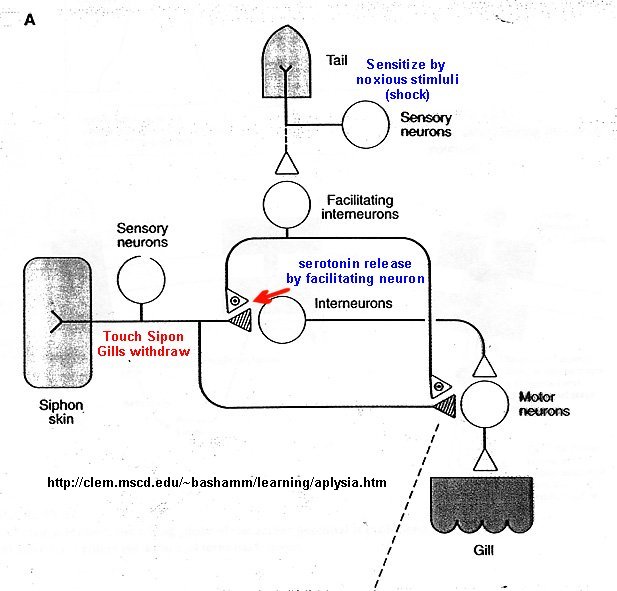
- Aplysis sensory cells in sensitization
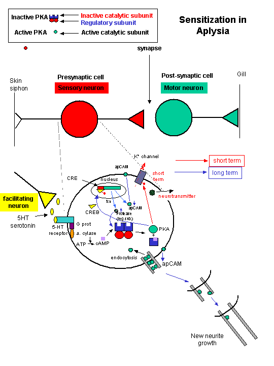
- Changes in Sensory Cells in Habituation
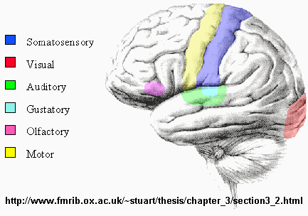
- Sensory regions of the brain
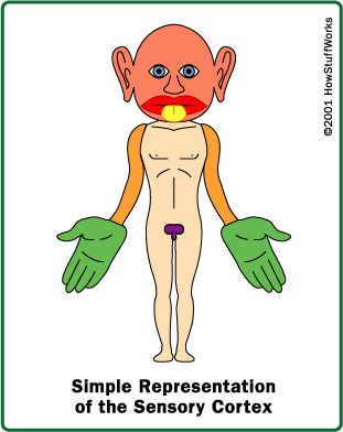
- Somatosensory Map - Homunculus
Capstone: Origin of Life
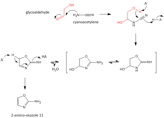
- Abiotic Synthesis of 2-amino-oxazole
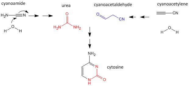
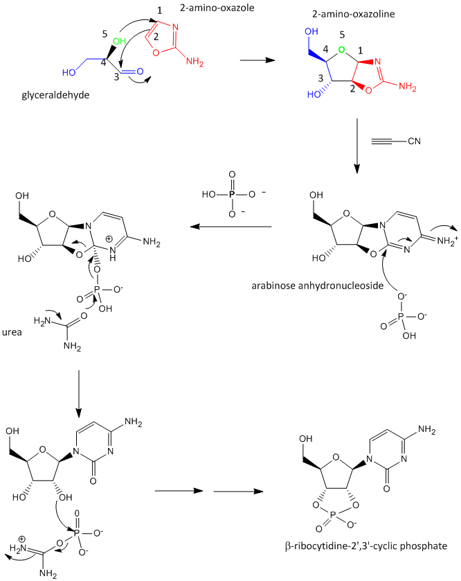
- Abiotic synthesis of a ribocytidine phosphate without condensation of a preformed ribose and cytosine
 Navigation
Return to Biochemistry Online Table of Contents

Biochemistry Online by Henry Jakubowski is licensed under a Creative Commons Attribution-NonCommercial 4.0 International License.